


 癌症的基本特征包括细胞增殖、血管生成、迁移、凋亡逃避机制和细胞永生等。找到癌症发生过程中这些通路的关键标记物和对应的抗体用于检测至关重要。
 为您推荐一个泛素化位点预测神器——泛素化分析工具,可以为您的蛋白的泛素化位点作出预测和评分。
 细胞自噬受体图形绘图工具为你的蛋白的细胞受体结合位点作出预测和评分,识别结合到自噬通路中的蛋白是非常重要的,便于让我们理解自噬在正常生理、病理过程中的作用,如发育、细胞分化、神经退化性疾病、压力条件下、感染和癌症。
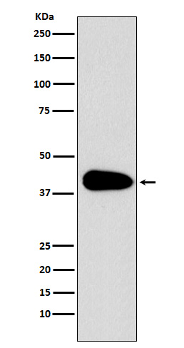
PAR6 Antibody
Rabbit mAb
- 产品详情
- 实验流程
Application
| WB, IP |
|---|---|
| Primary Accession | Q9NPB6 |
| Reactivity | Rat, Human, Mouse |
| Clonality | Monoclonal |
| Other Names | PAR6; PAR6C; TAX40; PAR-6A; TIP-40; PAR6alpha; |
| Isotype | Rabbit IgG |
| Host | Rabbit |
| Calculated MW | 37388 Da |
| Dilution | WB 1:500~1:2000 IP 1:50 |
|---|---|
| Purification | Affinity-chromatography |
| Immunogen | A synthesized peptide derived from human PAR6 |
| Description | Adapter protein involved in asymmetrical cell division and cell polarization processes. Probably involved in the formation of epithelial tight junctions. Association with PARD3 may prevent the interaction of PARD3 with F11R/JAM1, thereby preventing tight junction assembly. |
| Storage Condition and Buffer | Rabbit IgG in phosphate buffered saline , pH 7.4, 150mM NaCl, 0.02% sodium azide and 50% glycerol. Store at +4°C short term. Store at -20°C long term. Avoid freeze / thaw cycle. |
| Name | PARD6A |
|---|---|
| Synonyms | PAR6A |
| Function | Adapter protein involved in asymmetrical cell division and cell polarization processes. Probably involved in the formation of epithelial tight junctions. Association with PARD3 may prevent the interaction of PARD3 with F11R/JAM1, thereby preventing tight junction assembly. The PARD6-PARD3 complex links GTP-bound Rho small GTPases to atypical protein kinase C proteins (PubMed:10873802). Regulates centrosome organization and function. Essential for the centrosomal recruitment of key proteins that control centrosomal microtubule organization (PubMed:20719959). |
| Cellular Location | Cytoplasm. Cell membrane. Cell projection, ruffle. Cell junction, tight junction. Cytoplasm, cytoskeleton, microtubule organizing center, centrosome, centriolar satellite. Cytoplasm, cytoskeleton, microtubule organizing center, centrosome Note=Colocalizes with GTP-bound CDC42 or RAC1 at membrane ruffles and with PARD3 and PRKCI at epithelial tight junctions. Recruited to the centrosome by a microtubule and dynein-dynactin-dependent mechanism |
| Tissue Location | Expressed in pancreas, skeletal muscle, brain and heart. Weakly expressed in kidney and placenta |
Research Areas
For Research Use Only. Not For Use In Diagnostic Procedures.
Application Protocols
Provided below are standard protocols that you may find useful for product applications.
终于等到您。ABCEPTA(百远生物)抗体产品。
点击下方“我要评价 ”按钮提交您的反馈信息,您的反馈和评价是我们最宝贵的财富之一,
我们将在1-3个工作日内处理您的反馈信息。
如有疑问,联系:0512-88856768 tech-china@abcepta.com.


