


 癌症的基本特征包括细胞增殖、血管生成、迁移、凋亡逃避机制和细胞永生等。找到癌症发生过程中这些通路的关键标记物和对应的抗体用于检测至关重要。
 为您推荐一个泛素化位点预测神器——泛素化分析工具,可以为您的蛋白的泛素化位点作出预测和评分。
 细胞自噬受体图形绘图工具为你的蛋白的细胞受体结合位点作出预测和评分,识别结合到自噬通路中的蛋白是非常重要的,便于让我们理解自噬在正常生理、病理过程中的作用,如发育、细胞分化、神经退化性疾病、压力条件下、感染和癌症。
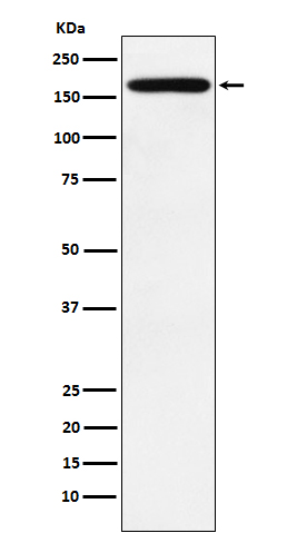
PODXL Antibody
Rabbit mAb
- 产品详情
- 实验流程
Application
| WB, IHC, IF, FC, ICC, IP, IHF |
|---|---|
| Primary Accession | O00592 |
| Reactivity | Human |
| Clonality | Monoclonal |
| Other Names | Gp2; Gp200; PC; PCLP; PCLP1; Pcx; Podocalyxin; Podocalyxin like; Podocalyxin-like protein 1; Podxl; |
| Isotype | Rabbit IgG |
| Host | Rabbit |
| Calculated MW | 58635 Da |
| Dilution | WB 1:500~1:2000 IHC 1:50~1:200 ICC/IF 1:100~1:500 IP 1:50 FC 1:100 |
|---|---|
| Purification | Affinity-chromatography |
| Immunogen | A synthesized peptide derived from human PODXL |
| Description | Involved in the regulation of both adhesion and cell morphology and cancer progression. Function as an anti-adhesive molecule that maintains an open filtration pathway between neighboring foot processes in the podocyte by charge repulsion. |
| Storage Condition and Buffer | Rabbit IgG in phosphate buffered saline , pH 7.4, 150mM NaCl, 0.02% sodium azide and 50% glycerol. Store at +4°C short term. Store at -20°C long term. Avoid freeze / thaw cycle. |
| Name | PODXL |
|---|---|
| Synonyms | PCLP, PCLP1 |
| Function | Involved in the regulation of both adhesion and cell morphology and cancer progression. Functions as an anti-adhesive molecule that maintains an open filtration pathway between neighboring foot processes in the podocyte by charge repulsion. Acts as a pro- adhesive molecule, enhancing the adherence of cells to immobilized ligands, increasing the rate of migration and cell-cell contacts in an integrin-dependent manner. Induces the formation of apical actin- dependent microvilli. Involved in the formation of a preapical plasma membrane subdomain to set up initial epithelial polarization and the apical lumen formation during renal tubulogenesis. Plays a role in cancer development and aggressiveness by inducing cell migration and invasion through its interaction with the actin-binding protein EZR. Affects EZR-dependent signaling events, leading to increased activities of the MAPK and PI3K pathways in cancer cells. |
| Cellular Location | Apical cell membrane. Cell projection, lamellipodium. Cell projection, filopodium. Cell projection, ruffle Cell projection, microvillus. Membrane raft. Membrane; Single-pass type I membrane protein. Note=In single attached epithelial cells is restricted to a preapical pole on the free plasma membrane whereas other apical and basolateral proteins are not yet polarized Colocalizes with NHERF2 at the apical plasma membrane during epithelial polarization. Colocalizes with NHERF1 at the trans-Golgi network (transiently) and at the apical plasma membrane. Its association with the membrane raft is transient. Colocalizes with actin filaments, EZR and NHERF1 in a punctate pattern at the apical cell surface where microvilli form. Colocalizes with EZR and NHERF2 at the apical cell membrane of glomerular epithelium cells (By similarity). Forms granular, punctuated pattern, forming patches, preferentially adopting a polar distribution, located on the migrating poles of the cell or forming clusters along the terminal ends of filipodia establishing contact with the endothelial cells. Colocalizes with the submembrane actin of lamellipodia, particularly associated with ruffles Colocalizes with vinculin at protrusions of cells. Colocalizes with ITGB1. Colocalizes with PARD3, PRKCI, EXOC5, OCLN, RAB11A and RAB8A in apical membrane initiation sites (AMIS) during the generation of apical surface and luminogenesis (By similarity). |
| Tissue Location | Glomerular epithelium cell (podocyte). |
Research Areas
For Research Use Only. Not For Use In Diagnostic Procedures.
Application Protocols
Provided below are standard protocols that you may find useful for product applications.
终于等到您。ABCEPTA(百远生物)抗体产品。
点击下方“我要评价 ”按钮提交您的反馈信息,您的反馈和评价是我们最宝贵的财富之一,
我们将在1-3个工作日内处理您的反馈信息。
如有疑问,联系:0512-88856768 tech-china@abcepta.com.
¥ 1,500.00
Cat# AP92071


