


 癌症的基本特征包括细胞增殖、血管生成、迁移、凋亡逃避机制和细胞永生等。找到癌症发生过程中这些通路的关键标记物和对应的抗体用于检测至关重要。
 为您推荐一个泛素化位点预测神器——泛素化分析工具,可以为您的蛋白的泛素化位点作出预测和评分。
 细胞自噬受体图形绘图工具为你的蛋白的细胞受体结合位点作出预测和评分,识别结合到自噬通路中的蛋白是非常重要的,便于让我们理解自噬在正常生理、病理过程中的作用,如发育、细胞分化、神经退化性疾病、压力条件下、感染和癌症。
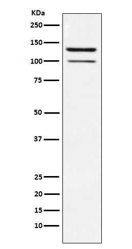
HIPK2 Antibody
Rabbit mAb
- 产品详情
- 实验流程
Application
| WB, IF, FC, ICC, IP |
|---|---|
| Primary Accession | Q9H2X6 |
| Reactivity | Human, Mouse |
| Clonality | Monoclonal |
| Other Names | hHIPk2; Hipk2; Nbak1; Stank; |
| Isotype | Rabbit IgG |
| Host | Rabbit |
| Calculated MW | 130966 Da |
| Dilution | WB 1:500~1:2000 ICC/IF 1:50~1:200 IP 1:30 FC 1:500 |
|---|---|
| Purification | Affinity-chromatography |
| Immunogen | A synthesized peptide derived from human HIPK2 |
| Description | Protein kinase acting as a corepressor of several transcription factors, including SMAD1 and POU4F1/Brn3a and probably NK homeodomain transcription factors. Inhibits cell growth and promotes apoptosis. Involved in transcriptional activation of TP53 and TP73. |
| Storage Condition and Buffer | Rabbit IgG in phosphate buffered saline , pH 7.4, 150mM NaCl, 0.02% sodium azide and 50% glycerol. Store at +4°C short term. Store at -20°C long term. Avoid freeze / thaw cycle. |
| Name | HIPK2 |
|---|---|
| Function | Serine/threonine-protein kinase involved in transcription regulation, p53/TP53-mediated cellular apoptosis and regulation of the cell cycle. Acts as a corepressor of several transcription factors, including SMAD1 and POU4F1/Brn3a and probably NK homeodomain transcription factors. Phosphorylates PDX1, ATF1, PML, p53/TP53, CREB1, CTBP1, CBX4, RUNX1, EP300, CTNNB1, HMGA1, ZBTB4 and DAZAP2. Inhibits cell growth and promotes apoptosis through the activation of p53/TP53 both at the transcription level and at the protein level (by phosphorylation and indirect acetylation). The phosphorylation of p53/TP53 may be mediated by a p53/TP53-HIPK2-AXIN1 complex. Involved in the response to hypoxia by acting as a transcriptional co-suppressor of HIF1A. Mediates transcriptional activation of TP73. In response to TGFB, cooperates with DAXX to activate JNK. Negative regulator through phosphorylation and subsequent proteasomal degradation of CTNNB1 and the antiapoptotic factor CTBP1. In the Wnt/beta-catenin signaling pathway acts as an intermediate kinase between MAP3K7/TAK1 and NLK to promote the proteasomal degradation of MYB. Phosphorylates CBX4 upon DNA damage and promotes its E3 SUMO-protein ligase activity. Activates CREB1 and ATF1 transcription factors by phosphorylation in response to genotoxic stress. In response to DNA damage, stabilizes PML by phosphorylation. PML, HIPK2 and FBXO3 may act synergically to activate p53/TP53-dependent transactivation. Promotes angiogenesis, and is involved in erythroid differentiation, especially during fetal liver erythropoiesis. Phosphorylation of RUNX1 and EP300 stimulates EP300 transcription regulation activity. Triggers ZBTB4 protein degradation in response to DNA damage. In response to DNA damage, phosphorylates DAZAP2 which localizes DAZAP2 to the nucleus, reduces interaction of DAZAP2 with HIPK2 and prevents DAZAP2-dependent ubiquitination of HIPK2 by E3 ubiquitin-protein ligase SIAH1 and subsequent proteasomal degradation (PubMed:33591310). Modulates HMGA1 DNA-binding affinity. In response to high glucose, triggers phosphorylation-mediated subnuclear localization shifting of PDX1. Involved in the regulation of eye size, lens formation and retinal lamination during late embryogenesis. |
| Cellular Location | Nucleus, PML body. Cytoplasm Cytoplasm, Stress granule Note=Concentrated in PML/POD/ND10 nuclear bodies. Small amounts are cytoplasmic |
| Tissue Location | Highly expressed in heart, muscle and kidney. Weakly expressed in a ubiquitous way. Down-regulated in several thyroid and breast tumors. |
Research Areas
For Research Use Only. Not For Use In Diagnostic Procedures.
Application Protocols
Provided below are standard protocols that you may find useful for product applications.
终于等到您。ABCEPTA(百远生物)抗体产品。
点击下方“我要评价 ”按钮提交您的反馈信息,您的反馈和评价是我们最宝贵的财富之一,
我们将在1-3个工作日内处理您的反馈信息。
如有疑问,联系:0512-88856768 tech-china@abcepta.com.
¥ 1,500.00
Cat# AP92137


