


 癌症的基本特征包括细胞增殖、血管生成、迁移、凋亡逃避机制和细胞永生等。找到癌症发生过程中这些通路的关键标记物和对应的抗体用于检测至关重要。
 为您推荐一个泛素化位点预测神器——泛素化分析工具,可以为您的蛋白的泛素化位点作出预测和评分。
 细胞自噬受体图形绘图工具为你的蛋白的细胞受体结合位点作出预测和评分,识别结合到自噬通路中的蛋白是非常重要的,便于让我们理解自噬在正常生理、病理过程中的作用,如发育、细胞分化、神经退化性疾病、压力条件下、感染和癌症。
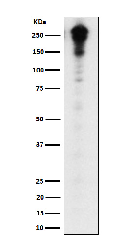
Desmoplakin Antibody
Rabbit mAb
- 产品详情
- 实验流程
Application
| WB, IF, FC, ICC |
|---|---|
| Primary Accession | P15924 |
| Reactivity | Rat, Human |
| Clonality | Monoclonal |
| Other Names | Desmoplakin I; Desmoplakin II; DPI; DPII; DSP; KPPS2; PPKS2; |
| Isotype | Rabbit IgG |
| Host | Rabbit |
| Calculated MW | 331774 Da |
| Dilution | WB 1:500~1:2000 ICC/IF 1:50~1:200 FC 1:50 |
|---|---|
| Purification | Affinity-chromatography |
| Immunogen | A synthesized peptide derived from human Desmoplakin |
| Description | Major high molecular weight protein of desmosomes. Involved in the organization of the desmosomal cadherin-plakoglobin complexes into discrete plasma membrane domains and in the anchoring of intermediate filaments to the desmosomes. |
| Storage Condition and Buffer | Rabbit IgG in phosphate buffered saline , pH 7.4, 150mM NaCl, 0.02% sodium azide and 50% glycerol. Store at +4°C short term. Store at -20°C long term. Avoid freeze / thaw cycle. |
| Name | DSP (HGNC:3052) |
|---|---|
| Function | A component of desmosome cell-cell junctions which are required for positive regulation of cellular adhesion (PubMed:25733715). Critical for cell-cell adhesion in early stage blastocysts and progression through proamniotic cavity formation (By similarity). Not required for preimplantation morphogenic process in blastocysts (By similarity). Required for keratin filament anchoring at the desmosome junction and subsequent organization of the keratin intermediate filament network within the cytoplasm (By similarity). Required for anchoring of desmosomes to the microtubule architecture, via its interaction with NIN (By similarity). Plays a key role in adhesion and organization of the dermal epithelial barrier (By similarity). Critical for the maintenance of the neural tube structure following formation and organization of the neuroepithelium (By similarity). Facilitates outgrowth and repair of motor neuron fibers in regenerating axons following injury, probably by promoting recruitment of a complex containing DSP, CDH2, VIM and JUP to the outgrowth tips (By similarity). Required for the normal formation of the heart, also required for development of vascular capillary structures and intact endothelial cell barriers (By similarity). Regulates profibrotic gene expression in cardiomyocytes via activation of the MAPK14/p38 MAPK signaling cascade and increase in TGFB1 protein abundance (By similarity). Maintains cardiac rhythmicity by ensuring correct cell- cell adhesion within the sinoatrial node, via stabilization of protein components of both desmosome and Gap junctions (By similarity). Involved in maintaining the protein stability and recruitment of GJA1 to functional gap junctions, via inhibition of KRAS-mediated MAPK1/MAPK3 phosphorylation of GJA1 (By similarity). Required for the survival and maintenance of germ cells in the gonads during embryonic development (By similarity). Binds to telomere DNA (via C-terminus) and acts to prevent telomere damage and maintain telomere length via its interaction with TRF2 (PubMed:31595153). |
| Cellular Location | Cell projection, axon {ECO:0000250|UniProtKB:E9Q557}. Cell junction, desmosome. Cell membrane {ECO:0000250|UniProtKB:E9Q557}. Cytoplasm. Nucleus Note=Localizes to desmosome precursor particles in the cytoplasm (PubMed:25208567). Localizes to the cytoplasm in undifferentiated keratinocytes however becomes localizes to both lateral and tricellular cell-cell contacts as differentiation progresses and as epithelial sheet formation completes (By similarity). Localizes to the interface of the cortical keratin network and endodermal cell periphery in developing embryos (By similarity). {ECO:0000250|UniProtKB:E9Q557, ECO:0000269|PubMed:25208567} |
| Tissue Location | Expressed in oral mucosa (at protein level) (PubMed:30479852). Expressed in arrector pili muscle (at protein level) (PubMed:29034528). Expressed in the heart in the heart (at protein level) (PubMed:18662195). [Isoform DPII]: Resides predominantly in tissues and cells of stratified origin |
Research Areas
For Research Use Only. Not For Use In Diagnostic Procedures.
Application Protocols
Provided below are standard protocols that you may find useful for product applications.
终于等到您。ABCEPTA(百远生物)抗体产品。
点击下方“我要评价 ”按钮提交您的反馈信息,您的反馈和评价是我们最宝贵的财富之一,
我们将在1-3个工作日内处理您的反馈信息。
如有疑问,联系:0512-88856768 tech-china@abcepta.com.


