


 癌症的基本特征包括细胞增殖、血管生成、迁移、凋亡逃避机制和细胞永生等。找到癌症发生过程中这些通路的关键标记物和对应的抗体用于检测至关重要。
 为您推荐一个泛素化位点预测神器——泛素化分析工具,可以为您的蛋白的泛素化位点作出预测和评分。
 细胞自噬受体图形绘图工具为你的蛋白的细胞受体结合位点作出预测和评分,识别结合到自噬通路中的蛋白是非常重要的,便于让我们理解自噬在正常生理、病理过程中的作用,如发育、细胞分化、神经退化性疾病、压力条件下、感染和癌症。
ICOS Antibody
Rabbit mAb
- 产品详情
- 实验流程
Application
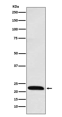
| WB, IF, FC, ICC |
|---|---|
| Primary Accession | Q9Y6W8 |
| Reactivity | Rat, Human, Mouse |
| Clonality | Monoclonal |
| Other Names | AILIM; CD278; CRP1; CVID1; ICOS; |
| Isotype | Rabbit IgG |
| Host | Rabbit |
| Calculated MW | 22625 Da |
| Dilution | WB 1:500~1:2000 ICC/IF 1:50~1:200 FC 1:50 |
|---|---|
| Purification | Affinity-chromatography |
| Immunogen | A synthesized peptide derived from human ICOS |
| Description | Enhances all basic T-cell responses to a foreign antigen, namely proliferation, secretion of lymphokines, up-regulation of molecules that mediate cell-cell interaction, and effective help for antibody secretion by B-cells. |
| Storage Condition and Buffer | Rabbit IgG in phosphate buffered saline , pH 7.4, 150mM NaCl, 0.02% sodium azide and 50% glycerol. Store at +4°C short term. Store at -20°C long term. Avoid freeze / thaw cycle. |
| Name | ICOS |
|---|---|
| Synonyms | AILIM |
| Function | Stimulatory receptor expressed in activated or antigen- experienced T-cells that plays an important role in the immune response (PubMed:9930702). Upon binding to its ligand ICOSL expressed on antigen presenting cells (APCs), delivers costimulatory signals that enhances all basic T-cell responses to a foreign antigen, namely proliferation, secretion of lymphokines including IL10, up-regulation of molecules that mediate cell-cell interaction, and effective help for antibody secretion by B-cells (PubMed:33033255). Also acts as a costimulatory receptor critical for the differentiation of T follicular regulatory cells upon immune challenges such as viral infection (PubMed:27135603). Mechanistically, potentiates TCR-induced calcium flux by augmenting PLCG1 activation and actin remodeling (By similarity). In addition, activates PI3K signaling pathways independently of calcium flux (PubMed:30523347). Essential both for efficient interaction between T and B-cells and for normal antibody responses to T-cell dependent antigens. Prevents the apoptosis of pre-activated T-cells. Plays a critical role in CD40-mediated class switching of immunoglobin isotypes (By similarity). |
| Cellular Location | [Isoform 1]: Cell membrane; Single-pass type I membrane protein |
| Tissue Location | Activated T-cells. Highly expressed on tonsillar T- cells, which are closely associated with B-cells in the apical light zone of germinal centers, the site of terminal B-cell maturation Expressed at lower levels in thymus, lung, lymph node and peripheral blood leukocytes. Expressed in the medulla of fetal and newborn thymus |
Research Areas
For Research Use Only. Not For Use In Diagnostic Procedures.
Application Protocols
Provided below are standard protocols that you may find useful for product applications.
终于等到您。ABCEPTA(百远生物)抗体产品。
点击下方“我要评价 ”按钮提交您的反馈信息,您的反馈和评价是我们最宝贵的财富之一,
我们将在1-3个工作日内处理您的反馈信息。
如有疑问,联系:0512-88856768 tech-china@abcepta.com.
¥ 1,500.00
Cat# AP92346


