


 癌症的基本特征包括细胞增殖、血管生成、迁移、凋亡逃避机制和细胞永生等。找到癌症发生过程中这些通路的关键标记物和对应的抗体用于检测至关重要。
 为您推荐一个泛素化位点预测神器——泛素化分析工具,可以为您的蛋白的泛素化位点作出预测和评分。
 细胞自噬受体图形绘图工具为你的蛋白的细胞受体结合位点作出预测和评分,识别结合到自噬通路中的蛋白是非常重要的,便于让我们理解自噬在正常生理、病理过程中的作用,如发育、细胞分化、神经退化性疾病、压力条件下、感染和癌症。
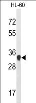
ATP5C1 Antibody (N-term)
Affinity Purified Rabbit Polyclonal Antibody (Pab)
- 产品详情
- 文献引用 : 1
- 实验流程
- 背景知识
Application
| WB, IHC-P, FC, E |
|---|---|
| Primary Accession | P36542 |
| Other Accession | P35435, Q91VR2, Q4R5B0, P05631 |
| Reactivity | Human |
| Predicted | Bovine, Monkey, Mouse, Rat |
| Host | Rabbit |
| Clonality | Polyclonal |
| Isotype | Rabbit IgG |
| Calculated MW | 32996 Da |
| Antigen Region | 40-67 aa |
| Gene ID | 509 |
|---|---|
| Other Names | ATP synthase subunit gamma, mitochondrial, F-ATPase gamma subunit, ATP5C1, ATP5C, ATP5CL1 |
| Target/Specificity | This ATP5C1 antibody is generated from rabbits immunized with a KLH conjugated synthetic peptide between 40-67 amino acids from the N-terminal region of human ATP5C1. |
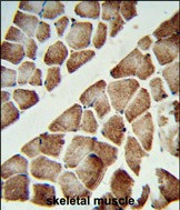
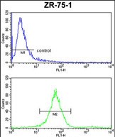
| Dilution | WB~~1:1000 IHC-P~~1:100~500 FC~~1:10~50 E~~Use at an assay dependent concentration. |
| Format | Purified polyclonal antibody supplied in PBS with 0.09% (W/V) sodium azide. This antibody is purified through a protein A column, followed by peptide affinity purification. |
| Storage | Maintain refrigerated at 2-8°C for up to 2 weeks. For long term storage store at -20°C in small aliquots to prevent freeze-thaw cycles. |
| Precautions | ATP5C1 Antibody (N-term) is for research use only and not for use in diagnostic or therapeutic procedures. |
| Name | ATP5F1C (HGNC:833) |
|---|---|
| Function | Subunit gamma, of the mitochondrial membrane ATP synthase complex (F(1)F(0) ATP synthase or Complex V) that produces ATP from ADP in the presence of a proton gradient across the membrane which is generated by electron transport complexes of the respiratory chain (PubMed:37244256). ATP synthase complex consist of a soluble F(1) head domain - the catalytic core - and a membrane F(1) domain - the membrane proton channel (PubMed:37244256). These two domains are linked by a central stalk rotating inside the F(1) region and a stationary peripheral stalk (PubMed:37244256). During catalysis, ATP synthesis in the catalytic domain of F(1) is coupled via a rotary mechanism of the central stalk subunits to proton translocation (Probable). In vivo, can only synthesize ATP although its ATP hydrolase activity can be activated artificially in vitro (By similarity). With the central stalk subunit delta, is essential for the biogenesis of F(1) catalytic part of the ATP synthase complex namely in the formation of F1 assembly intermediate (PubMed:29499186). |
| Cellular Location | Mitochondrion inner membrane {ECO:0000250|UniProtKB:P05631}; Peripheral membrane protein {ECO:0000250|UniProtKB:P05631}; Matrix side {ECO:0000250|UniProtKB:P05631} |
| Tissue Location | Isoform Heart is expressed specifically in the heart and skeletal muscle, which require rapid energy supply. Isoform Liver is expressed in the brain, liver and kidney. Isoform Heart and Isoform Liver are expressed in the skin, intestine, stomach and aorta |
For Research Use Only. Not For Use In Diagnostic Procedures.

Provided below are standard protocols that you may find useful for product applications.
BACKGROUND
ATP5C1 encodes a subunit of mitochondrial ATP synthase. Mitochondrial ATP synthase catalyzes ATP synthesis, utilizing an electrochemical gradient of protons across the inner membrane during oxidative phosphorylation. ATP synthase is composed of two linked multi-subunit complexes: the soluble catalytic core, F1, and the membrane-spanning component, Fo, comprising the proton channel. The catalytic portion of mitochondrial ATP synthase consists of 5 different subunits (alpha, beta, gamma, delta, and epsilon) assembled with a stoichiometry of 3 alpha, 3 beta, and a single representative of the other 3.
REFERENCES
Wheeler,H.E., et.al, PLoS Genet. 5 (10), E1000685 (2009)
Wang,L., et.al, Cancer Epidemiol. Biomarkers Prev. 17 (12), 3558-3566 (2008)
终于等到您。ABCEPTA(百远生物)抗体产品。
点击下方“我要评价 ”按钮提交您的反馈信息,您的反馈和评价是我们最宝贵的财富之一,
我们将在1-3个工作日内处理您的反馈信息。
如有疑问,联系:0512-88856768 tech-china@abcepta.com.


