


 癌症的基本特征包括细胞增殖、血管生成、迁移、凋亡逃避机制和细胞永生等。找到癌症发生过程中这些通路的关键标记物和对应的抗体用于检测至关重要。
 为您推荐一个泛素化位点预测神器——泛素化分析工具,可以为您的蛋白的泛素化位点作出预测和评分。
 细胞自噬受体图形绘图工具为你的蛋白的细胞受体结合位点作出预测和评分,识别结合到自噬通路中的蛋白是非常重要的,便于让我们理解自噬在正常生理、病理过程中的作用,如发育、细胞分化、神经退化性疾病、压力条件下、感染和癌症。
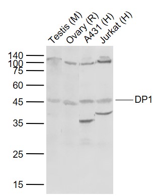
DP1 Rabbit pAb
DP1 Rabbit pAb
- 产品详情
- 实验流程
- 背景知识
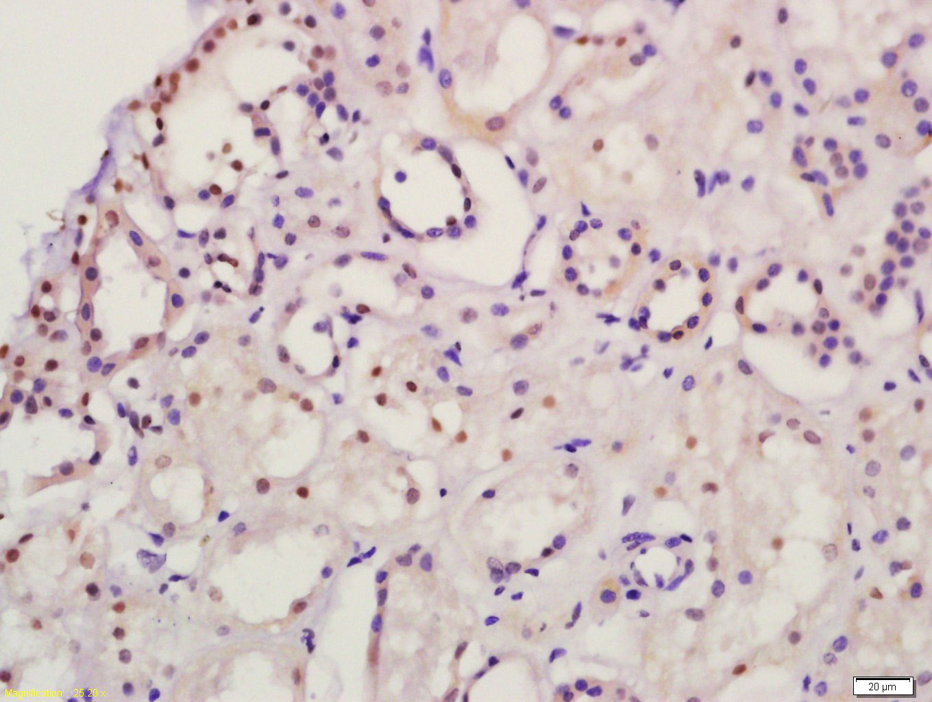
Application
| WB, IHC-P, IHC-F, IF |
|---|---|
| Primary Accession | Q14186 |
| Reactivity | Human, Mouse, Rat |
| Predicted | Chicken, Dog, Horse, Rabbit |
| Host | Rabbit |
| Clonality | Polyclonal |
| Calculated MW | 45070 Da |
| Physical State | Liquid |
| Immunogen | KLH conjugated synthetic peptide derived from human TFDP1 |
| Epitope Specificity | 221-320/410 |
| Isotype | IgG |
| Purity | affinity purified by Protein A |
| Buffer | 0.01M TBS (pH7.4) with 1% BSA, 0.02% Proclin300 and 50% Glycerol. |
| SUBCELLULAR LOCATION | Nucleus. |
| SIMILARITY | Belongs to the E2F/DP family. |
| SUBUNIT | Component of the E2F/DP transcription factor complex. Forms heterodimers with E2F family members. The complex can interact with hypophosphorylated retinoblastoma protein RB1 and related proteins (RBL1 and RBL2) that inhibit the E2F transactivation domain. This repression involves recruitment of histone deacetylase (HDAC). During the cell cycle, from mid-to-late G1 phase, RB family members become phosphorylated, detach from the DRTF1/E2F complex to render E2F transcriptionally active. Viral oncoproteins, notably E1A, T-antigen and HPV E7, are capable of sequestering RB protein, thus releasing the active complex. Part of the E2F6.com-1 complex in G0 phase is composed of E2F6, MGA, MAX, TFDP1, CBX3, BAT8, EUHMTASE1, RING1, RNF2, MBLR, L3MBTL2 YAF2. Component of the DREAM complex (also named LINC complex) at least composed of E2F4, E2F5, LIN9, LIN37, LIN52, LIN54, MYBL1, MYBL2, RBL1, RBL2, RBBP4, TFDP1 and TFDP2. The complex exists in quiescent cells where it represses cell cycle-dependent genes. It dissociates in S phase when LIN9, LIN37, LIN52 and LIN54 form a subcomplex that binds to MYBL2. |
| Post-translational modifications | Phosphorylation by E2F-1-bound cyclin A-CDK2, in the S phase, inhibits E2F-mediated DNA binding and transactivation. |
| Important Note | This product as supplied is intended for research use only, not for use in human, therapeutic or diagnostic applications. |
| Background Descriptions | This gene encodes a member of a family of transcription factors that heterodimerize with E2F proteins to enhance their DNA-binding activity and promote transcription from E2F target genes. The encoded protein functions as part of this complex to control the transcriptional activity of numerous genes involved in cell cycle progression from G1 to S phase. Alternative splicing results in multiple transcript variants. Pseudogenes of this gene are found on chromosomes 1, 15, and X.[provided by RefSeq, Jan 2009] |
| Gene ID | 7027 |
|---|---|
| Other Names | Transcription factor Dp-1, DRTF1-polypeptide 1, DRTF1, E2F dimerization partner 1, TFDP1, DP1 |
| Target/Specificity | Highest levels in muscle. Also expressed in brain, placenta, liver and kidney. Lower levels in lung and pancreas. |
| Dilution | WB=1:500-2000,IHC-P=1:100-500,IHC-F=1:100-500,IF=1:100-500 |
| Storage | Store at -20 °C for one year. Avoid repeated freeze/thaw cycles. When reconstituted in sterile pH 7.4 0.01M PBS or diluent of antibody the antibody is stable for at least two weeks at 2-4 °C. |
| Name | TFDP1 |
|---|---|
| Synonyms | DP1 |
| Function | Can stimulate E2F-dependent transcription. Binds DNA cooperatively with E2F family members through the E2 recognition site, 5'-TTTC[CG]CGC-3', found in the promoter region of a number of genes whose products are involved in cell cycle regulation or in DNA replication (PubMed:7739537, PubMed:8405995). The E2F1:DP complex appears to mediate both cell proliferation and apoptosis. Blocks adipocyte differentiation by repressing CEBPA binding to its target gene promoters (PubMed:20176812). |
| Cellular Location | Nucleus {ECO:0000250|UniProtKB:Q08639}. Cytoplasm {ECO:0000250|UniProtKB:Q08639}. Note=Shuttles between the cytoplasm and nucleus and translocates into the nuclear compartment upon heterodimerization with E2F1. {ECO:0000250|UniProtKB:Q08639} |
| Tissue Location | Highest levels in muscle. Also expressed in brain, placenta, liver and kidney. Lower levels in lung and pancreas. Not detected in heart |
For Research Use Only. Not For Use In Diagnostic Procedures.
Provided below are standard protocols that you may find useful for product applications.
BACKGROUND
This gene encodes a member of a family of transcription factors that heterodimerize with E2F proteins to enhance their DNA-binding activity and promote transcription from E2F target genes. The encoded protein functions as part of this complex to control the transcriptional activity of numerous genes involved in cell cycle progression from G1 to S phase. Alternative splicing results in multiple transcript variants. Pseudogenes of this gene are found on chromosomes 1, 15, and X.[provided by RefSeq, Jan 2009]
终于等到您。ABCEPTA(百远生物)抗体产品。
点击下方“我要评价 ”按钮提交您的反馈信息,您的反馈和评价是我们最宝贵的财富之一,
我们将在1-3个工作日内处理您的反馈信息。
如有疑问,联系:0512-88856768 tech-china@abcepta.com.


