


 癌症的基本特征包括细胞增殖、血管生成、迁移、凋亡逃避机制和细胞永生等。找到癌症发生过程中这些通路的关键标记物和对应的抗体用于检测至关重要。
 为您推荐一个泛素化位点预测神器——泛素化分析工具,可以为您的蛋白的泛素化位点作出预测和评分。
 细胞自噬受体图形绘图工具为你的蛋白的细胞受体结合位点作出预测和评分,识别结合到自噬通路中的蛋白是非常重要的,便于让我们理解自噬在正常生理、病理过程中的作用,如发育、细胞分化、神经退化性疾病、压力条件下、感染和癌症。
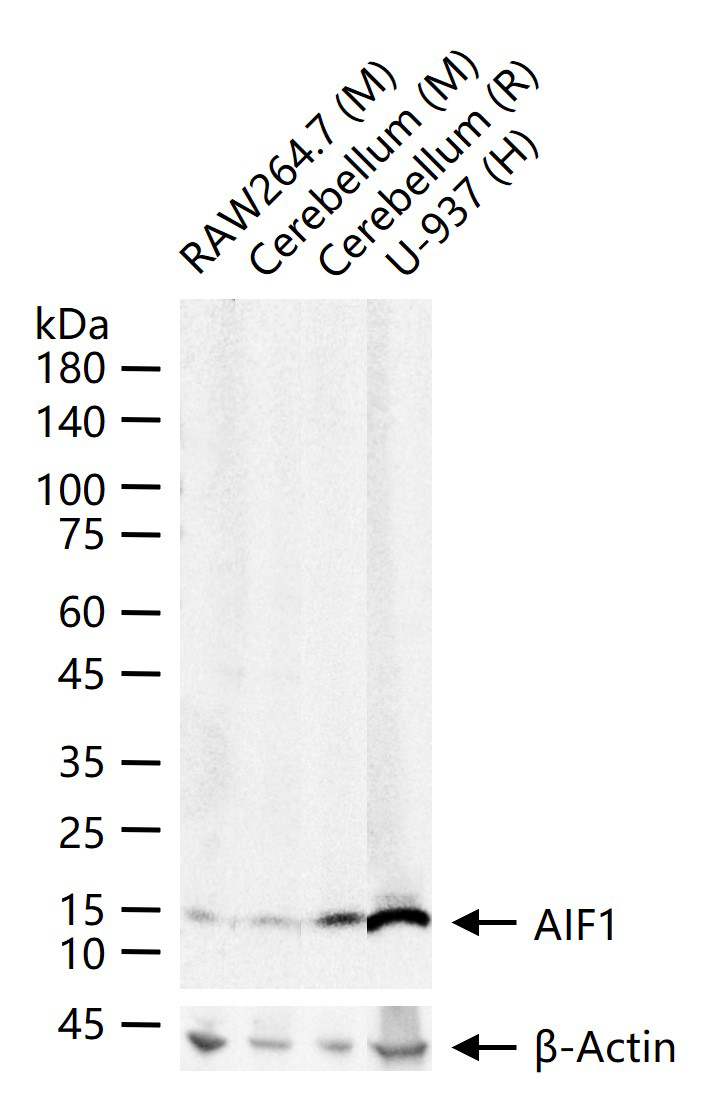
AIF1 / Iba1 Rabbit pAb
AIF1 / Iba1 Rabbit pAb
- 产品详情
- 实验流程
- 背景知识
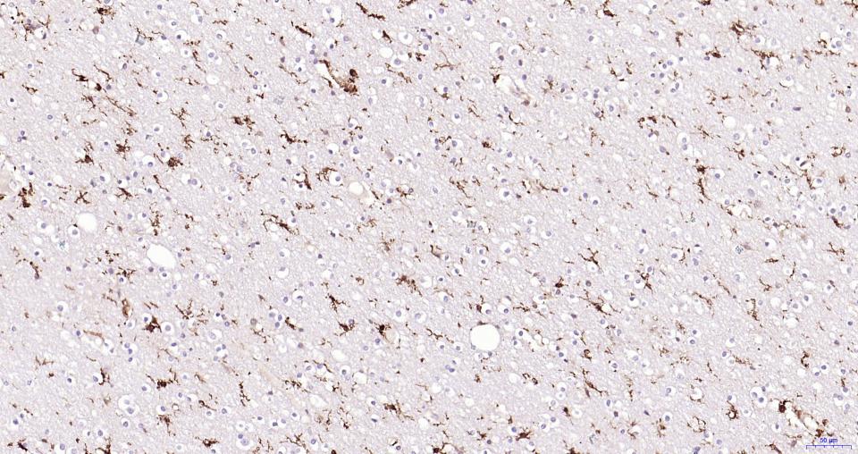
Application
| WB, IHC-P, IHC-F, IF |
|---|---|
| Primary Accession | P55008 |
| Reactivity | Human, Mouse, Rat |
| Host | Rabbit |
| Clonality | Polyclonal |
| Calculated MW | 16703 Da |
| Physical State | Liquid |
| Immunogen | KLH conjugated synthetic peptide derived from human Iba1 |
| Epitope Specificity | 51-147/147 |
| Isotype | IgG |
| Purity | affinity purified by Protein A |
| Buffer | 0.01M TBS (pH7.4) with 1% BSA, 0.02% Proclin300 and 50% Glycerol. |
| SUBCELLULAR LOCATION | Cytoplasm, cytoskeleton. Cell projection, ruffle membrane; Peripheral membrane protein; Cytoplasmic side. Note=Associated with the actin cytoskeleton at membrane ruffles and at sites of phagocytosis. |
| SIMILARITY | Contains 2 EF-hand domains. |
| SUBUNIT | Homodimer (Potential). Monomer. Interacts with LCP1. |
| Important Note | This product as supplied is intended for research use only, not for use in human, therapeutic or diagnostic applications. |
| Background Descriptions | Allograft Inflammatory Factor-1 (AIF1)or ionized calcium-binding adaptor molecule 1 (Iba1) is expressed selectively in microglia/macrophages and is a Ca2+-binding peptide produced by activated monocytes and microglial cells. It has been suggested that AIF1 expression is associated with chronic inflammatory processes. AIF1 is expressed by activated monocytes and might participate in a variety of pathogenic processes in the mammalian brain and in chronic transplant rejection. It has been shown to be expressed early and persistently in chronically rejecting cardiac allografts but not in cardiac syngrafts and host hearts. |
| Gene ID | 199 |
|---|---|
| Other Names | Allograft inflammatory factor 1, AIF-1, Ionized calcium-binding adapter molecule 1, Protein G1, AIF1, G1, IBA1 |
| Target/Specificity | Detected in T-lymphocytes and peripheral blood mononuclear cells. |
| Dilution | WB=1:500-1000,IHC-P=1:100-500,IHC-F=1:100-500,IF=1:100-500 |
| Storage | Store at -20 °C for one year. Avoid repeated freeze/thaw cycles. When reconstituted in sterile pH 7.4 0.01M PBS or diluent of antibody the antibody is stable for at least two weeks at 2-4 °C. |
| Name | AIF1 |
|---|---|
| Synonyms | G1, IBA1 |
| Function | Actin-binding protein that enhances membrane ruffling and RAC activation. Enhances the actin-bundling activity of LCP1. Binds calcium. Plays a role in RAC signaling and in phagocytosis. May play a role in macrophage activation and function. Promotes the proliferation of vascular smooth muscle cells and of T-lymphocytes. Enhances lymphocyte migration. Plays a role in vascular inflammation. |
| Cellular Location | Cytoplasm, cytoskeleton {ECO:0000250|UniProtKB:O70200}. Cell projection, ruffle membrane {ECO:0000250|UniProtKB:O70200}; Peripheral membrane protein {ECO:0000250|UniProtKB:O70200}; Cytoplasmic side {ECO:0000250|UniProtKB:O70200}. Cell projection, phagocytic cup {ECO:0000250|UniProtKB:O70200}. Note=Associated with the actin cytoskeleton at membrane ruffles and at sites of phagocytosis {ECO:0000250|UniProtKB:O70200} |
| Tissue Location | Detected in T-lymphocytes and peripheral blood mononuclear cells. |
For Research Use Only. Not For Use In Diagnostic Procedures.
Provided below are standard protocols that you may find useful for product applications.
BACKGROUND
Allograft Inflammatory Factor-1 (AIF1)or ionized calcium-binding adaptor molecule 1 (Iba1) is expressed selectively in microglia/macrophages and is a Ca2+-binding peptide produced by activated monocytes and microglial cells. It has been suggested that AIF1 expression is associated with chronic inflammatory processes. AIF1 is expressed by activated monocytes and might participate in a variety of pathogenic processes in the mammalian brain and in chronic transplant rejection. It has been shown to be expressed early and persistently in chronically rejecting cardiac allografts but not in cardiac syngrafts and host hearts.
终于等到您。ABCEPTA(百远生物)抗体产品。
点击下方“我要评价 ”按钮提交您的反馈信息,您的反馈和评价是我们最宝贵的财富之一,
我们将在1-3个工作日内处理您的反馈信息。
如有疑问,联系:0512-88856768 tech-china@abcepta.com.


