


 癌症的基本特征包括细胞增殖、血管生成、迁移、凋亡逃避机制和细胞永生等。找到癌症发生过程中这些通路的关键标记物和对应的抗体用于检测至关重要。
 为您推荐一个泛素化位点预测神器——泛素化分析工具,可以为您的蛋白的泛素化位点作出预测和评分。
 细胞自噬受体图形绘图工具为你的蛋白的细胞受体结合位点作出预测和评分,识别结合到自噬通路中的蛋白是非常重要的,便于让我们理解自噬在正常生理、病理过程中的作用,如发育、细胞分化、神经退化性疾病、压力条件下、感染和癌症。
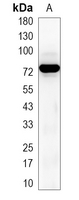
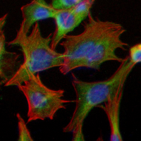
Recombinant Anti-TAK1 Rabbit mAb
- 产品详情
- 实验流程
Application 
| WB, IHC, FC, IF/IC |
|---|---|
| Primary Accession | O43318 |
| Reactivity | Human, Mouse, Rat |
| Host | Rabbit |
| Clonality | Monoclonal |
| Calculated MW | 67196 Da |
| Gene ID | 6885 |
|---|---|
| Other Names | TAK1; Mitogen-activated protein kinase kinase kinase 7; Transforming growth factor-beta-activated kinase 1; TGF-beta-activated kinase 1 |
| Target/Specificity | KLH-conjugated synthetic peptide encompassing a sequence within human TAK1 protein. The exact sequence is proprietary. |
| Dilution | WB~~WB (1/500 - 1/1000) IHC~~1:100~500 FC~~1:10~50 IF/IC~~N/A |
| Format | Liquid in PBS, pH 7.3, 50% glycerol, 0.05% BSA, and 0.05% Proclin300. |
| Storage | Store at -20 °C.Stable for 12 months from date of receipt |
For Research Use Only. Not For Use In Diagnostic Procedures.
| Name | MAP3K7 {ECO:0000303|PubMed:28397838, ECO:0000312|HGNC:HGNC:6859} |
|---|---|
| Function | Serine/threonine kinase which acts as an essential component of the MAP kinase signal transduction pathway (PubMed:10094049, PubMed:11460167, PubMed:12589052, PubMed:16845370, PubMed:16893890, PubMed:21512573, PubMed:8663074, PubMed:9079627). Plays an important role in the cascades of cellular responses evoked by changes in the environment (PubMed:10094049, PubMed:11460167, PubMed:12589052, PubMed:16845370, PubMed:16893890, PubMed:21512573, PubMed:8663074, PubMed:9079627). Mediates signal transduction of TRAF6, various cytokines including interleukin-1 (IL-1), transforming growth factor- beta (TGFB), TGFB-related factors like BMP2 and BMP4, toll-like receptors (TLR), tumor necrosis factor receptor CD40 and B-cell receptor (BCR) (PubMed:16893890, PubMed:9079627). Once activated, acts as an upstream activator of the MKK/JNK signal transduction cascade and the p38 MAPK signal transduction cascade through the phosphorylation and activation of several MAP kinase kinases like MAP2K1/MEK1, MAP2K3/MKK3, MAP2K6/MKK6 and MAP2K7/MKK7 (PubMed:11460167, PubMed:8663074). These MAP2Ks in turn activate p38 MAPKs and c-jun N- terminal kinases (JNKs); both p38 MAPK and JNK pathways control the transcription factors activator protein-1 (AP-1) (PubMed:11460167, PubMed:12589052, PubMed:8663074). Independently of MAP2Ks and p38 MAPKs, acts as a key activator of NF-kappa-B by promoting activation of the I-kappa-B-kinase (IKK) core complex (PubMed:12589052, PubMed:8663074). Mechanistically, recruited to polyubiquitin chains of RIPK2 and IKBKG/NEMO via TAB2/MAP3K7IP2 and TAB3/MAP3K7IP3, and catalyzes phosphorylation and activation of IKBKB/IKKB component of the IKK complex, leading to NF-kappa-B activation (PubMed:10094049, PubMed:11460167). In osmotic stress signaling, plays a major role in the activation of MAPK8/JNK1, but not that of NF-kappa-B (PubMed:16893890). Promotes TRIM5 capsid-specific restriction activity (PubMed:21512573). Phosphorylates RIPK1 at 'Ser-321' which positively regulates RIPK1 interaction with RIPK3 to promote necroptosis but negatively regulates RIPK1 kinase activity and its interaction with FADD to mediate apoptosis (By similarity). Phosphorylates STING1 in response to cGAMP-activation, promoting association between STEEP1 and STING1 and STING1 translocation to COPII vesicles (PubMed:37832545). |
| Cellular Location | Cytoplasm. Cell membrane; Peripheral membrane protein; Cytoplasmic side. Note=Although the majority of MAP3K7/TAK1 is found in the cytosol, when complexed with TAB1/MAP3K7IP1 and TAB2/MAP3K7IP2, it is also localized at the cell membrane |
| Tissue Location | Isoform 1A is the most abundant in ovary, skeletal muscle, spleen and blood mononuclear cells. Isoform 1B is highly expressed in brain, kidney and small intestine. Isoform 1C is the major form in prostate. Isoform 1D is the less abundant form |
Research Areas
Application Protocols
Provided below are standard protocols that you may find useful for product applications.

终于等到您。ABCEPTA(百远生物)抗体产品。
点击下方“我要评价 ”按钮提交您的反馈信息,您的反馈和评价是我们最宝贵的财富之一,
我们将在1-3个工作日内处理您的反馈信息。
如有疑问,联系:0512-88856768 tech-china@abcepta.com.
¥ 1,250.00
Cat# AP95088


