


 癌症的基本特征包括细胞增殖、血管生成、迁移、凋亡逃避机制和细胞永生等。找到癌症发生过程中这些通路的关键标记物和对应的抗体用于检测至关重要。
 为您推荐一个泛素化位点预测神器——泛素化分析工具,可以为您的蛋白的泛素化位点作出预测和评分。
 细胞自噬受体图形绘图工具为你的蛋白的细胞受体结合位点作出预测和评分,识别结合到自噬通路中的蛋白是非常重要的,便于让我们理解自噬在正常生理、病理过程中的作用,如发育、细胞分化、神经退化性疾病、压力条件下、感染和癌症。
Anti-PDPK1 Antibody
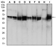
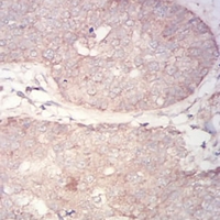
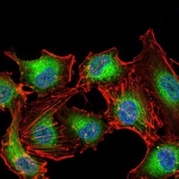
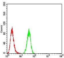
- 产品详情
- 实验流程
Application 
| WB, IHC, FC, IF/IC |
|---|---|
| Primary Accession | O15530 |
| Reactivity | Human, Mouse, Rat, Mk |
| Host | Mouse |
| Clonality | Monoclonal |
| Calculated MW | 63152 Da |
| Gene ID | 5170 |
|---|---|
| Other Names | PDK1; 3-phosphoinositide-dependent protein kinase 1; hPDK1 |
| Target/Specificity | Recombinant fusion protein of human PDPK1 expressed in E. Coli |
| Dilution | WB~~WB (1/500 - 1/1000) IHC~~1:100~500 FC~~FC (1/100 - 1/200) IF/IC~~N/A |
| Format | Mouse IgG1. Liquid in PBS, pH 7.3, 30% glycerol, and 0.01% sodium azide. |
| Storage | Store at -20 °C.Stable for 12 months from date of receipt |
For Research Use Only. Not For Use In Diagnostic Procedures.
| Name | PDPK1 |
|---|---|
| Synonyms | PDK1 |
| Function | Serine/threonine kinase which acts as a master kinase, phosphorylating and activating a subgroup of the AGC family of protein kinases (PubMed:10226025, PubMed:10480933, PubMed:10995762, PubMed:12167717, PubMed:14585963, PubMed:14604990, PubMed:16207722, PubMed:16251192, PubMed:17327236, PubMed:17371830, PubMed:18835241, PubMed:9094314, PubMed:9368760, PubMed:9445476, PubMed:9445477, PubMed:9707564, PubMed:9768361). Its targets include: protein kinase B (PKB/AKT1, PKB/AKT2, PKB/AKT3), p70 ribosomal protein S6 kinase (RPS6KB1), p90 ribosomal protein S6 kinase (RPS6KA1, RPS6KA2 and RPS6KA3), cyclic AMP-dependent protein kinase (PRKACA), protein kinase C (PRKCD and PRKCZ), serum and glucocorticoid-inducible kinase (SGK1, SGK2 and SGK3), p21-activated kinase-1 (PAK1), TSSK3, protein kinase PKN (PKN1 and PKN2) (PubMed:10226025, PubMed:10480933, PubMed:10995762, PubMed:12167717, PubMed:14585963, PubMed:14604990, PubMed:16207722, PubMed:16251192, PubMed:17327236, PubMed:17371830, PubMed:18835241, PubMed:9094314, PubMed:9368760, PubMed:9445476, PubMed:9707564, PubMed:9768361). Plays a central role in the transduction of signals from insulin by providing the activating phosphorylation to PKB/AKT1, thus propagating the signal to downstream targets controlling cell proliferation and survival, as well as glucose and amino acid uptake and storage (PubMed:10226025, PubMed:12167717, PubMed:9094314). Negatively regulates the TGF-beta-induced signaling by: modulating the association of SMAD3 and SMAD7 with TGF-beta receptor, phosphorylating SMAD2, SMAD3, SMAD4 and SMAD7, preventing the nuclear translocation of SMAD3 and SMAD4 and the translocation of SMAD7 from the nucleus to the cytoplasm in response to TGF-beta (PubMed:17327236). Activates PPARG transcriptional activity and promotes adipocyte differentiation (By similarity). Activates the NF-kappa-B pathway via phosphorylation of IKKB (PubMed:16207722). The tyrosine phosphorylated form is crucial for the regulation of focal adhesions by angiotensin II (PubMed:14585963). Controls proliferation, survival, and growth of developing pancreatic cells (By similarity). Participates in the regulation of Ca(2+) entry and Ca(2+)-activated K(+) channels of mast cells (By similarity). Essential for the motility of vascular endothelial cells (ECs) and is involved in the regulation of their chemotaxis (PubMed:17371830). Plays a critical role in cardiac homeostasis by serving as a dual effector for cell survival and beta-adrenergic response (By similarity). Plays an important role during thymocyte development by regulating the expression of key nutrient receptors on the surface of pre-T cells and mediating Notch-induced cell growth and proliferative responses (By similarity). Provides negative feedback inhibition to toll-like receptor-mediated NF-kappa-B activation in macrophages (By similarity). |
| Cellular Location | Cytoplasm. Nucleus. Cell membrane; Peripheral membrane protein. Cell junction, focal adhesion. Note=Tyrosine phosphorylation seems to occur only at the cell membrane. Translocates to the cell membrane following insulin stimulation by a mechanism that involves binding to GRB14 and INSR. SRC and HSP90 promote its localization to the cell membrane. Its nuclear localization is dependent on its association with PTPN6 and its phosphorylation at Ser- 396. Restricted to the nucleus in neuronal cells while in non-neuronal cells it is found in the cytoplasm. The Ser-241 phosphorylated form is distributed along the perinuclear region in neuronal cells while in non-neuronal cells it is found in both the nucleus and the cytoplasm IGF1 transiently increases phosphorylation at Ser-241 of neuronal PDPK1, resulting in its translocation to other cellular compartments The tyrosine-phosphorylated form colocalizes with PTK2B in focal adhesions after angiotensin II stimulation |
| Tissue Location | Appears to be expressed ubiquitously. The Tyr-9 phosphorylated form is markedly increased in diseased tissue compared with normal tissue from lung, liver, colon and breast |
Research Areas
Application Protocols
Provided below are standard protocols that you may find useful for product applications.

终于等到您。ABCEPTA(百远生物)抗体产品。
点击下方“我要评价 ”按钮提交您的反馈信息,您的反馈和评价是我们最宝贵的财富之一,
我们将在1-3个工作日内处理您的反馈信息。
如有疑问,联系:0512-88856768 tech-china@abcepta.com.
¥ 1,250.00
Cat# AP95998


