


 癌症的基本特征包括细胞增殖、血管生成、迁移、凋亡逃避机制和细胞永生等。找到癌症发生过程中这些通路的关键标记物和对应的抗体用于检测至关重要。
 为您推荐一个泛素化位点预测神器——泛素化分析工具,可以为您的蛋白的泛素化位点作出预测和评分。
 细胞自噬受体图形绘图工具为你的蛋白的细胞受体结合位点作出预测和评分,识别结合到自噬通路中的蛋白是非常重要的,便于让我们理解自噬在正常生理、病理过程中的作用,如发育、细胞分化、神经退化性疾病、压力条件下、感染和癌症。
COX6A2 Antibody (Center)
Affinity Purified Rabbit Polyclonal Antibody (Pab)
- 产品详情
- 文献引用 : 1
- 实验流程
- 背景知识
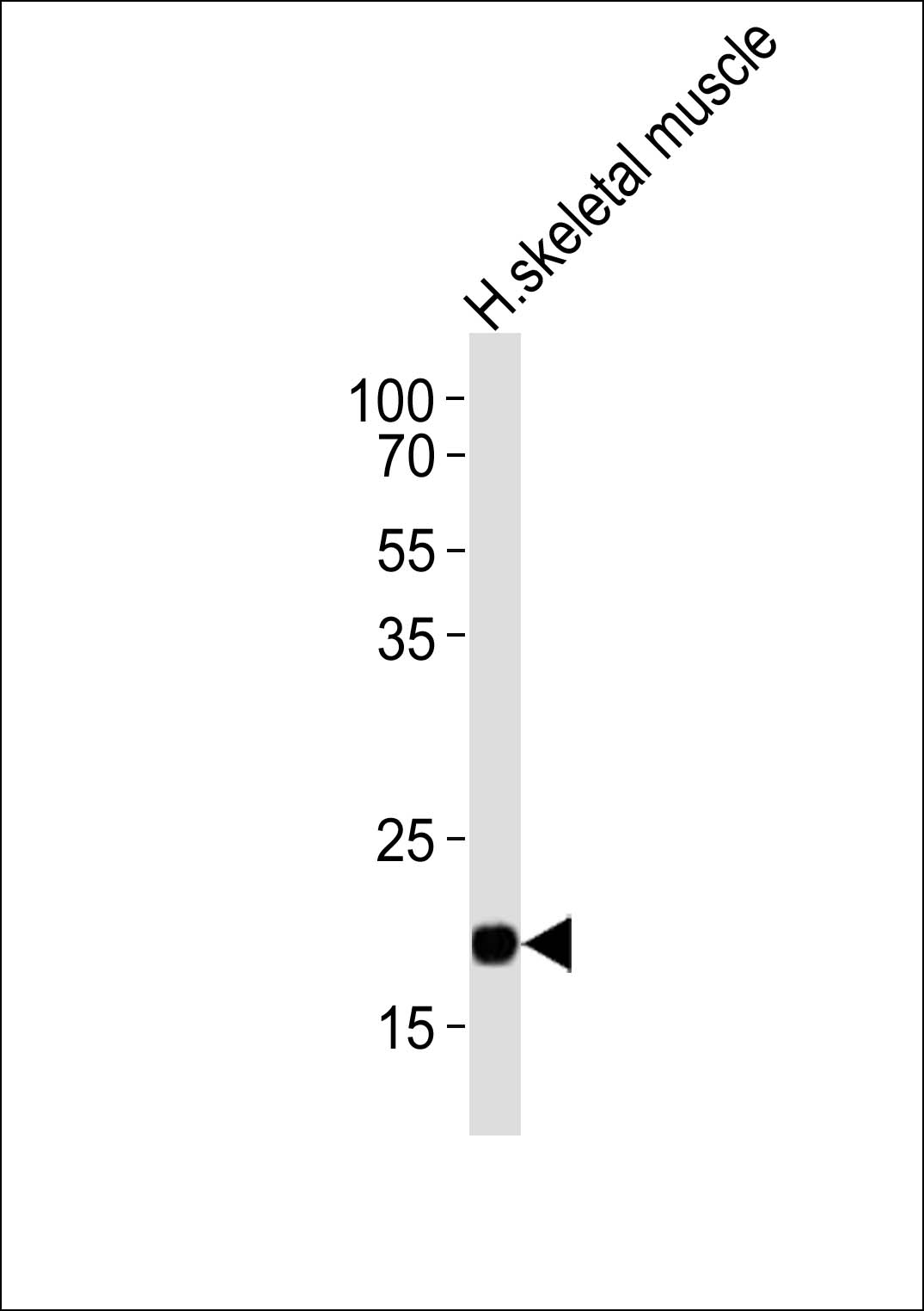
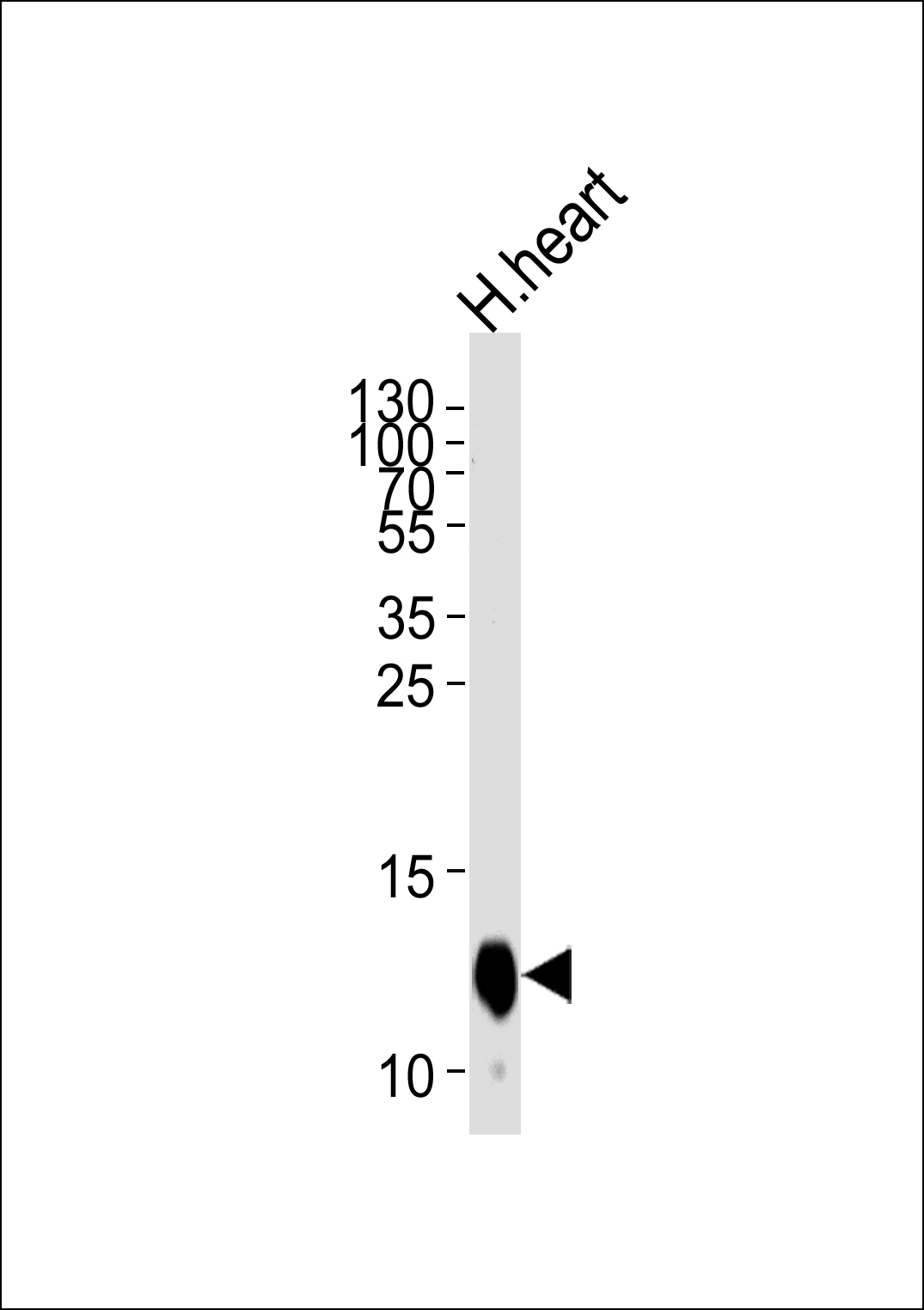
Application
| WB, IHC-P, E |
|---|---|
| Primary Accession | Q02221 |
| Reactivity | Human |
| Host | Rabbit |
| Clonality | Polyclonal |
| Isotype | Rabbit IgG |
| Calculated MW | 10815 Da |
| Antigen Region | 37-66 aa |
| Gene ID | 1339 |
|---|---|
| Other Names | Cytochrome c oxidase subunit 6A2, mitochondrial, Cytochrome c oxidase polypeptide VIa-heart, COXVIAH, Cytochrome c oxidase subunit VIA-muscle, COX VIa-M, COX6A2, COX6A, COX6AH |
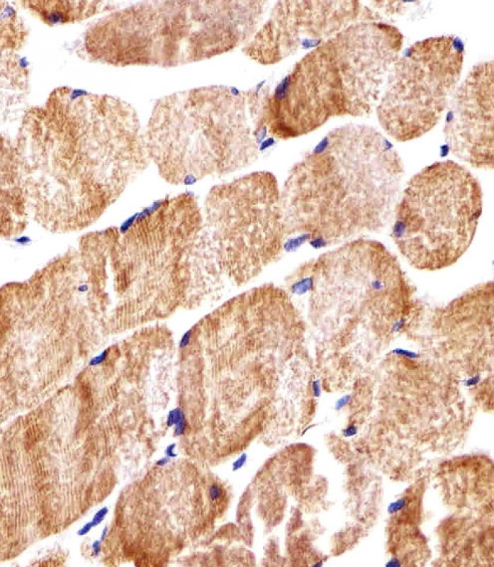
| Target/Specificity | This COX6A2 antibody is generated from rabbits immunized with a KLH conjugated synthetic peptide between 37-66 amino acids from the Central region of human COX6A2. |
| Dilution | WB~~1:1000 IHC-P~~1:100~500 E~~Use at an assay dependent concentration. |
| Format | Purified polyclonal antibody supplied in PBS with 0.09% (W/V) sodium azide. This antibody is purified through a protein A column, followed by peptide affinity purification. |
| Storage | Maintain refrigerated at 2-8°C for up to 2 weeks. For long term storage store at -20°C in small aliquots to prevent freeze-thaw cycles. |
| Precautions | COX6A2 Antibody (Center) is for research use only and not for use in diagnostic or therapeutic procedures. |
| Name | COX6A2 |
|---|---|
| Synonyms | COX6A, COX6AH |
| Function | Component of the cytochrome c oxidase, the last enzyme in the mitochondrial electron transport chain which drives oxidative phosphorylation. The respiratory chain contains 3 multisubunit complexes succinate dehydrogenase (complex II, CII), ubiquinol- cytochrome c oxidoreductase (cytochrome b-c1 complex, complex III, CIII) and cytochrome c oxidase (complex IV, CIV), that cooperate to transfer electrons derived from NADH and succinate to molecular oxygen, creating an electrochemical gradient over the inner membrane that drives transmembrane transport and the ATP synthase. Cytochrome c oxidase is the component of the respiratory chain that catalyzes the reduction of oxygen to water. Electrons originating from reduced cytochrome c in the intermembrane space (IMS) are transferred via the dinuclear copper A center (CU(A)) of subunit 2 and heme A of subunit 1 to the active site in subunit 1, a binuclear center (BNC) formed by heme A3 and copper B (CU(B)). The BNC reduces molecular oxygen to 2 water molecules unsing 4 electrons from cytochrome c in the IMS and 4 protons from the mitochondrial matrix. Plays a role in the assembly and stabilization of complex IV (PubMed:31155743). |
| Cellular Location | Mitochondrion inner membrane {ECO:0000250|UniProtKB:P07471}; Single-pass membrane protein {ECO:0000250|UniProtKB:P07471} |
| Tissue Location | Expressed specifically in heart and muscle (PubMed:31155743). Not detected in brain, colon, spleen, kidney, liver, lung and pancreas (PubMed:31155743). |
For Research Use Only. Not For Use In Diagnostic Procedures.

Provided below are standard protocols that you may find useful for product applications.
BACKGROUND
Cytochrome c oxidase (COX), the terminal enzyme of the mitochondrial respiratory chain, catalyzes the electron transfer from reduced cytochrome c to oxygen. It is a heteromeric complex consisting of 3 catalytic subunits encoded by mitochondrial genes and multiple structural subunits encoded by nuclear genes. The mitochondrially-encoded subunits function in electron transfer, and the nuclear-encoded subunits may be involved in the regulation and assembly of the complex. This nuclear gene encodes polypeptide 2 (heart/muscle isoform) of subunit VIa, and polypeptide 2 is present only in striated muscles. Polypeptide 1 (liver isoform) of subunit VIa is encoded by a different gene, and is found in all non-muscle tissues. These two polypeptides share 66% amino acid sequence identity.
REFERENCES
Bachman, N.J., et al. Genomics 42(1):146-151(1997)
Lanfranchi, G., et al. Genome Res. 6(1):35-42(1996)
终于等到您。ABCEPTA(百远生物)抗体产品。
点击下方“我要评价 ”按钮提交您的反馈信息,您的反馈和评价是我们最宝贵的财富之一,
我们将在1-3个工作日内处理您的反馈信息。
如有疑问,联系:0512-88856768 tech-china@abcepta.com.


