


 癌症的基本特征包括细胞增殖、血管生成、迁移、凋亡逃避机制和细胞永生等。找到癌症发生过程中这些通路的关键标记物和对应的抗体用于检测至关重要。
 为您推荐一个泛素化位点预测神器——泛素化分析工具,可以为您的蛋白的泛素化位点作出预测和评分。
 细胞自噬受体图形绘图工具为你的蛋白的细胞受体结合位点作出预测和评分,识别结合到自噬通路中的蛋白是非常重要的,便于让我们理解自噬在正常生理、病理过程中的作用,如发育、细胞分化、神经退化性疾病、压力条件下、感染和癌症。
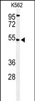
CBAA1 Antibody (N-term)
Affinity Purified Rabbit Polyclonal Antibody (Pab)
- 产品详情
- 实验流程
Application
| WB, E |
|---|---|
| Primary Accession | Q9BPX6 |
| Other Accession | Q4R518 |
| Reactivity | Human |
| Predicted | Monkey |
| Host | Rabbit |
| Clonality | Polyclonal |
| Isotype | Rabbit IgG |
| Calculated MW | 54351 Da |
| Antigen Region | 52-81 aa |
| Gene ID | 10367 |
|---|---|
| Other Names | Calcium uptake protein 1, mitochondrial, Atopy-related autoantigen CALC, ara CALC, Calcium-binding atopy-related autoantigen 1, Hom s 4, MICU1, CALC, CBARA1 |
| Target/Specificity | This CBAA1 antibody is generated from rabbits immunized with a KLH conjugated synthetic peptide between 52-81 amino acids from the N-terminal region of human CBAA1. |
| Dilution | WB~~1:1000 E~~Use at an assay dependent concentration. |
| Format | Purified polyclonal antibody supplied in PBS with 0.09% (W/V) sodium azide. This antibody is purified through a protein A column, followed by peptide affinity purification. |
| Storage | Maintain refrigerated at 2-8°C for up to 2 weeks. For long term storage store at -20°C in small aliquots to prevent freeze-thaw cycles. |
| Precautions | CBAA1 Antibody (N-term) is for research use only and not for use in diagnostic or therapeutic procedures. |
| Name | MICU1 {ECO:0000303|PubMed:20693986, ECO:0000312|HGNC:HGNC:1530} |
|---|---|
| Function | Calcium sensor of the mitochondrial calcium uniporter (MCU) channel, which senses calcium level via its EF-hand domains (PubMed:20693986, PubMed:23101630, PubMed:23747253, PubMed:24313810, PubMed:24332854, PubMed:24503055, PubMed:24560927, PubMed:26341627, PubMed:26903221, PubMed:27099988, PubMed:28615291, PubMed:30454562, PubMed:30638448, PubMed:32494073, PubMed:32667285, PubMed:32762847, PubMed:32790952, PubMed:34463251, PubMed:36206740, PubMed:37036971, PubMed:37126688). MICU1 and MICU2 (or MICU3) form a disulfide-linked heterodimer that stimulates and inhibits MCU activity, depending on the concentration of calcium (PubMed:24560927, PubMed:26903221, PubMed:28615291, PubMed:32148862, PubMed:32494073, PubMed:32667285, PubMed:32762847, PubMed:32790952, PubMed:36206740, PubMed:37036971, PubMed:37126688). At low calcium levels, MICU1 occludes the pore of the MCU channel, preventing mitochondrial calcium uptake (PubMed:32494073, PubMed:32667285, PubMed:32762847, PubMed:37036971, PubMed:37126688). At higher calcium levels, calcium-binding to MICU1 and MICU2 (or MICU3) induces a conformational change that weakens MCU-MICU1 interactions and moves the MICU1-MICU2 heterodimer away from the pore, allowing calcium permeation through the MCU channel (PubMed:32494073, PubMed:32667285, PubMed:32762847). Also required to protect against manganese toxicity by preventing manganese uptake by MCU: mechanistically, manganese- binding to its EF-hand domains does not induce any conformational change, maintaining MCU pore occlusion (PubMed:30082385, PubMed:30403999). Also acts as a barrier for inhibitors of the MCU channel, such as ruthenium red or its derivative Ru360 (PubMed:37244260). Acts as a regulator of mitochondrial cristae structure independently of its ability to regulate the mitochondrial calcium uniporter channel (PubMed:31427612, PubMed:37098122). Regulates glucose-dependent insulin secretion in pancreatic beta-cells by regulating mitochondrial calcium uptake (PubMed:22904319). Induces T- helper 1-mediated autoreactivity, which is accompanied by the release of IFNG (PubMed:16002733). |
| Cellular Location | Mitochondrion intermembrane space. Mitochondrion inner membrane. Note=Recruited to the mitochondrial inner membrane by EMRE/SMDT1 (PubMed:30454562). Also localizes to mitochondrial cristae junctions (PubMed:31427612) |
| Tissue Location | Expressed in epithelial cell lines. Strongly expressed in epidermal keratinocytes and dermal endothelial cells |
Research Areas
For Research Use Only. Not For Use In Diagnostic Procedures.
Application Protocols
Provided below are standard protocols that you may find useful for product applications.
REFERENCES
Bordicchia, M., et al. Metab. Clin. Exp. (2009) In press :
Grupe, A., et al. Am. J. Hum. Genet. 78(1):78-88(2006)
Natter, S., et al. FASEB J. 12(14):1559-1569(1998)
终于等到您。ABCEPTA(百远生物)抗体产品。
点击下方“我要评价 ”按钮提交您的反馈信息,您的反馈和评价是我们最宝贵的财富之一,
我们将在1-3个工作日内处理您的反馈信息。
如有疑问,联系:0512-88856768 tech-china@abcepta.com.
¥ 1,250.00
Cat# AP9682a


