


 癌症的基本特征包括细胞增殖、血管生成、迁移、凋亡逃避机制和细胞永生等。找到癌症发生过程中这些通路的关键标记物和对应的抗体用于检测至关重要。
 为您推荐一个泛素化位点预测神器——泛素化分析工具,可以为您的蛋白的泛素化位点作出预测和评分。
 细胞自噬受体图形绘图工具为你的蛋白的细胞受体结合位点作出预测和评分,识别结合到自噬通路中的蛋白是非常重要的,便于让我们理解自噬在正常生理、病理过程中的作用,如发育、细胞分化、神经退化性疾病、压力条件下、感染和癌症。
TMEM173 Antibody (C-term) 精选
Affinity Purified Rabbit Polyclonal Antibody (Pab)
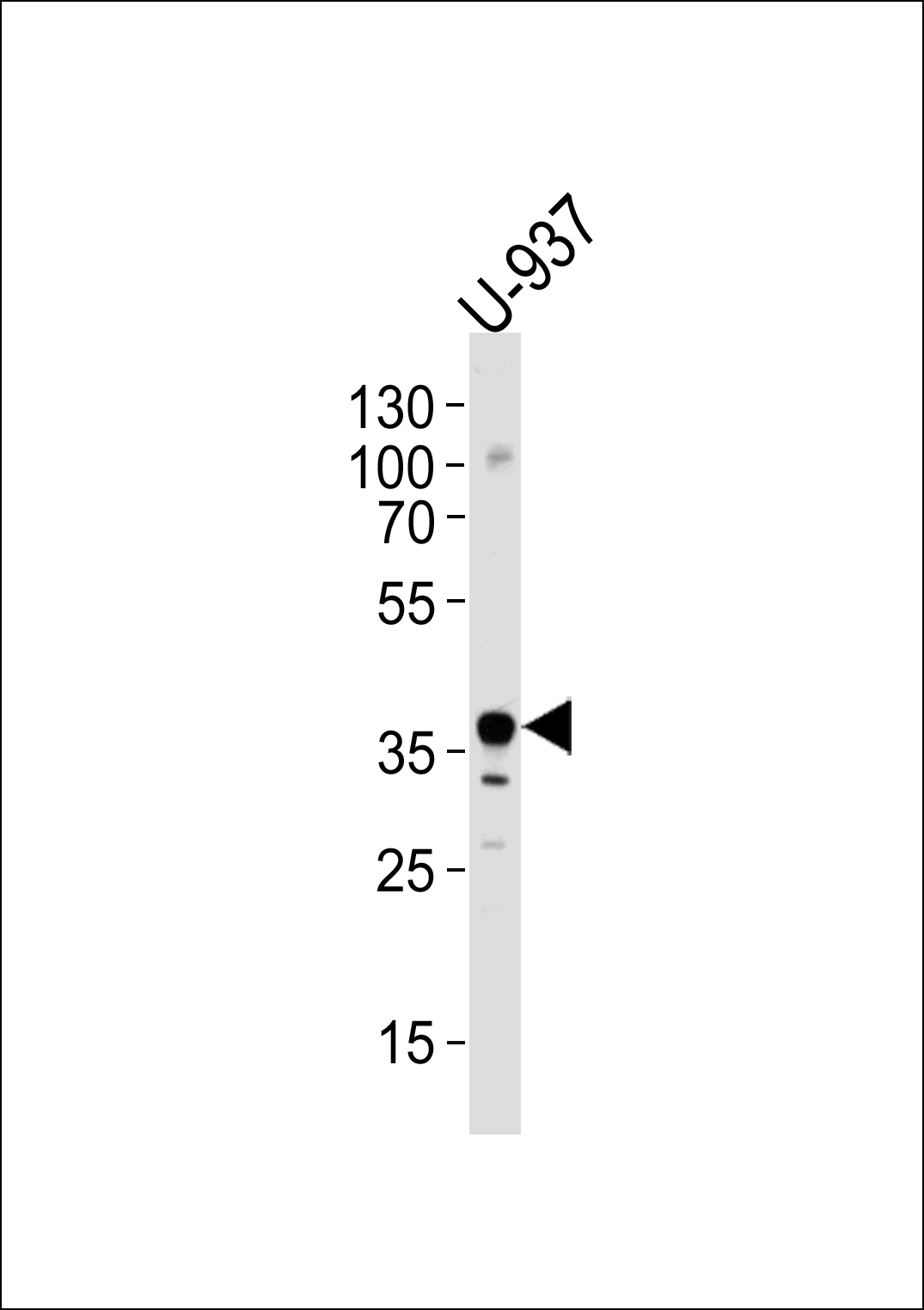
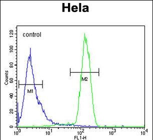
- 产品详情
- 实验流程
- 背景知识
Application 
| FC, WB, E |
|---|---|
| Primary Accession | Q86WV6 |
| Reactivity | Human |
| Predicted | Mouse, Rat, Canine, Rabbit |
| Host | Rabbit |
| Clonality | Polyclonal |
| Isotype | Rabbit IgG |
| Calculated MW | 42193 Da |
| Antigen Region | 311-340 aa |
| Gene ID | 340061 |
|---|---|
| Other Names | Stimulator of interferon genes protein, hSTING, Endoplasmic reticulum interferon stimulator, ERIS, Mediator of IRF3 activation, hMITA, Transmembrane protein 173, TMEM173, ERIS, MITA, STING |
| Target/Specificity | This TMEM173 antibody is generated from rabbits immunized with a KLH conjugated synthetic peptide between 311-340 amino acids from the C-terminal region of human TMEM173. |
| Dilution | FC~~1:10~50 WB~~1:1000 E~~Use at an assay dependent concentration. |
| Format | Purified polyclonal antibody supplied in PBS with 0.09% (W/V) sodium azide. This antibody is purified through a protein A column, followed by peptide affinity purification. |
| Storage | Maintain refrigerated at 2-8°C for up to 2 weeks. For long term storage store at -20°C in small aliquots to prevent freeze-thaw cycles. |
| Precautions | TMEM173 Antibody (C-term) is for research use only and not for use in diagnostic or therapeutic procedures. |
For Research Use Only. Not For Use In Diagnostic Procedures.
| Name | STING1 (HGNC:27962) |
|---|---|
| Function | Key innate immune signaling adapter that promotes the production of type I interferon (IFN-alpha and IFN-beta) in response to the presence of DNA from bacteria and viruses in the cytosol (PubMed:18724357, PubMed:18818105, PubMed:19433799, PubMed:19776740, PubMed:23027953, PubMed:23747010, PubMed:23910378, PubMed:25704810, PubMed:27801882, PubMed:29973723, PubMed:30842659, PubMed:31992625, PubMed:32926474, PubMed:35045565, PubMed:35388221, PubMed:36808561, PubMed:37086726, PubMed:37832545, PubMed:39255680, PubMed:39947179). Innate immune response is triggered by non-CpG double-stranded DNA from viruses and bacteria delivered to the cytoplasm, which induces production of cyclic dinucleotides that bind and activate STING1: STING1 recognizes and binds cyclic di-GMP (c-di-GMP), a second messenger produced by bacteria, cyclic UMP-AMP (2',3'-cUAMP), and cyclic GMP-AMP (cGAMP), a messenger produced by CGAS in response to DNA in the cytosol (PubMed:21947006, PubMed:23258412, PubMed:23707065, PubMed:23722158, PubMed:23747010, PubMed:23910378, PubMed:26229117, PubMed:26300263, PubMed:30842659, PubMed:35388221, PubMed:37086726, PubMed:37379839). Upon binding to c-di-GMP, cUAMP or cGAMP, STING1 oligomerizes and buds from the endoplasmic reticulum into COPII vesicles, which then form the endoplasmic reticulum-Golgi intermediate compartment (ERGIC) (PubMed:30842662, PubMed:41639452, PubMed:41639454, PubMed:41887218). It is then phosphorylated by TBK1 on the pLxIS motif, leading to recruitment and subsequent activation of the transcription factor IRF3 to induce expression of type I interferon and exert a potent antiviral state (PubMed:22394562, PubMed:25636800, PubMed:29973723, PubMed:30643259, PubMed:30842653, PubMed:32926474, PubMed:35045565, PubMed:35388221, PubMed:38917796). Also involved in intercellular immune signaling: cross-activated by 2',3'-cGAMP previously generated in virus-infected cells, triggering type I interferon signaling in macrophages and uninfected neighboring cells to propagate and amplify the antiviral immune response (PubMed:24077100, PubMed:31992625). In addition to promote the production of type I interferon, cGAS-STING signaling also activates the NF-kappa-B signaling: mechanistically, STING1 recruits TRAF6, leading to degradation of the NF-kappa-B inhibitor and subsequent translocation of NF-kappa-B into the nucleus (PubMed:32268090, PubMed:38917796, PubMed:39262777, PubMed:40973797, PubMed:41747053). Activation of NF- kappa-B signaling takes place in the endolysosome and is independent of type I interferon response via direct activation by RNA viruses (PubMed:40973797, PubMed:41747053). Independently of type I interferon production, plays a direct role in autophagy (PubMed:30568238, PubMed:30842662, PubMed:32926474). The ERGIC serves as the membrane source for WIPI2 recruitment and LC3 lipidation, leading to formation of autophagosomes that target cytosolic DNA or DNA viruses for degradation by the lysosome (PubMed:30842662). Promotes autophagy by acting as a proton channel that directs proton efflux from the Golgi to facilitate MAP1LC3B/LC3B lipidation (PubMed:37535724, PubMed:39947179). The autophagy- and interferon-inducing activities can be uncoupled and autophagy induction is independent of TBK1 phosphorylation (PubMed:30568238, PubMed:30842662). Autophagy is also triggered upon infection by bacteria: following c-di-GMP-binding, which is produced by live Gram-positive bacteria, promotes reticulophagy (By similarity). The proton channel activity also regulates lysosome biogenesis by activating transcription factors TFEB and TFE3, driving the expression of lysosome-related genes: activated STING1 in post-Golgi vesicles induces proton efflux and lipidation of GABARAP, sequestering the FLCN- FNIP complex and blocking mTORC1-dependent inhibition of TFEB and TFE3 (PubMed:39423796, PubMed:39689715). May be involved in transduction of apoptotic signals via its association with the major histocompatibility complex class II (MHC-II) (By similarity). |
| Cellular Location | Endoplasmic reticulum-Golgi intermediate compartment membrane; Multi-pass membrane protein {ECO:0000255, ECO:0000269|PubMed:32690950, ECO:0000269|PubMed:41639452}. Endoplasmic reticulum membrane; Multi-pass membrane protein {ECO:0000255, ECO:0000269|PubMed:30842659, ECO:0000269|PubMed:32690950}. Cytoplasm, perinuclear region. Golgi apparatus membrane; Multi-pass membrane protein. Golgi apparatus, trans-Golgi network membrane; Multi-pass membrane protein. Cytoplasmic vesicle, autophagosome membrane; Multi-pass membrane protein. Endosome membrane; Multi-pass membrane protein. Lysosome membrane; Multi-pass membrane protein. Mitochondrion outer membrane; Multi-pass membrane protein. Cell membrane {ECO:0000250|UniProtKB:Q3TBT3}; Multi-pass membrane protein. Note=Localizes to the endoplasmic reticulum when inactive (PubMed:19433799, PubMed:29694889, PubMed:30842653, PubMed:30842659). Following activation by cyclic dinucleotides, such as cGAMP, translocates from the endoplasmic reticulum to the endoplasmic reticulum-Golgi intermediate compartment (ERGIC) in a COPII vesicles- dependent process, where the kinase TBK1 is recruited (PubMed:19433799, PubMed:29694889, PubMed:30842653, PubMed:30842659, PubMed:37832545) Translocation to the ERGIC compartment is also dependent on cholesterol and phosphoinositide, such as phosphatidylinositol 3,5-bisphosphate (PtdIns(3,5)P2), which directly bind STING1 at the interface between dimers and promote STING1 homooligomerization (PubMed:41639452, PubMed:41639454). STING1-containing ERGIC serves as a membrane source for LC3 lipidation, which is a key step in autophagosome biogenesis (PubMed:30842662). Activates NF-kappa-B signaling when localized to the endolysosome compartment (PubMed:40973797). Localizes in the lysosome membrane in a TMEM203-dependent manner (By similarity) {ECO:0000250|UniProtKB:Q3TBT3, ECO:0000269|PubMed:19433799, ECO:0000269|PubMed:29694889, ECO:0000269|PubMed:30842653, ECO:0000269|PubMed:30842659, ECO:0000269|PubMed:30842662, ECO:0000269|PubMed:32690950, ECO:0000269|PubMed:37832545, ECO:0000269|PubMed:40973797, ECO:0000269|PubMed:41639452, ECO:0000269|PubMed:41639454} |
| Tissue Location | Ubiquitously expressed (PubMed:18724357, PubMed:18818105). Expressed in skin endothelial cells, alveolar type 2 pneumocytes, bronchial epithelium and alveolar macrophages (PubMed:25029335). |
Provided below are standard protocols that you may find useful for product applications.
BACKGROUND
Acts as a facilitator of innate immune signaling. Able to activate both NF-kappa-B and IRF3 transcription pathways to induce expression of type I interferon (IFN-alpha and IFN-beta) and exert a potent anti-viral state following expression. May be involved in translocon function, the translocon possibly being able to influence the induction of type I interferons. May be involved in transduction of apoptotic signals via its association with the major histocompatibility complex class II (MHC-II). Mediates death signaling via activation of the extracellular signal-regulated kinase (ERK) pathway.
REFERENCES
Sun, W., et al. Proc. Natl. Acad. Sci. U.S.A. 106(21):8653-8658(2009)
Zhong, B., et al. Immunity 30(3):397-407(2009)
Graubert, T.A., et al. PLoS ONE 4 (2), E4583 (2009)

终于等到您。ABCEPTA(百远生物)抗体产品。
点击下方“我要评价 ”按钮提交您的反馈信息,您的反馈和评价是我们最宝贵的财富之一,
我们将在1-3个工作日内处理您的反馈信息。
如有疑问,联系:0512-88856768 tech-china@abcepta.com.


