


 癌症的基本特征包括细胞增殖、血管生成、迁移、凋亡逃避机制和细胞永生等。找到癌症发生过程中这些通路的关键标记物和对应的抗体用于检测至关重要。
 为您推荐一个泛素化位点预测神器——泛素化分析工具,可以为您的蛋白的泛素化位点作出预测和评分。
 细胞自噬受体图形绘图工具为你的蛋白的细胞受体结合位点作出预测和评分,识别结合到自噬通路中的蛋白是非常重要的,便于让我们理解自噬在正常生理、病理过程中的作用,如发育、细胞分化、神经退化性疾病、压力条件下、感染和癌症。
DNAJC6 Antibody (Center)
Affinity Purified Rabbit Polyclonal Antibody (Pab)
- 产品详情
- 实验流程
- 背景知识
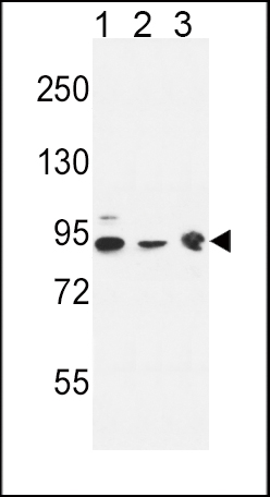
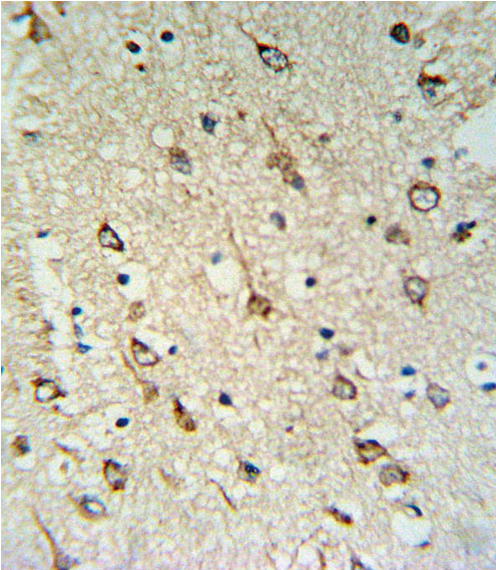
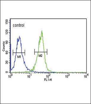
Application
| WB, IHC-P, FC, E |
|---|---|
| Primary Accession | O75061 |
| Other Accession | Q80TZ3 |
| Reactivity | Human, Rat, Mouse |
| Host | Rabbit |
| Clonality | Polyclonal |
| Isotype | Rabbit IgG |
| Calculated MW | 99997 Da |
| Antigen Region | 254-281 aa |
| Gene ID | 9829 |
|---|---|
| Other Names | Putative tyrosine-protein phosphatase auxilin, DnaJ homolog subfamily C member 6, DNAJC6, KIAA0473 |
| Target/Specificity | This DNAJC6 antibody is generated from rabbits immunized with a KLH conjugated synthetic peptide between 254-281 amino acids from the Central region of human DNAJC6. |
| Dilution | WB~~1:1000 IHC-P~~1:100~500 FC~~1:10~50 E~~Use at an assay dependent concentration. |
| Format | Purified polyclonal antibody supplied in PBS with 0.09% (W/V) sodium azide. This antibody is purified through a protein A column, followed by peptide affinity purification. |
| Storage | Maintain refrigerated at 2-8°C for up to 2 weeks. For long term storage store at -20°C in small aliquots to prevent freeze-thaw cycles. |
| Precautions | DNAJC6 Antibody (Center) is for research use only and not for use in diagnostic or therapeutic procedures. |
| Name | DNAJC6 (HGNC:15469) |
|---|---|
| Function | May act as a protein phosphatase and/or a lipid phosphatase. Co-chaperone that recruits HSPA8/HSC70 to clathrin-coated vesicles (CCVs) and promotes the ATP-dependent dissociation of clathrin from CCVs and participates in clathrin-mediated endocytosis of synaptic vesicles and their recycling and also in intracellular trafficking (PubMed:18489706). Firstly, binds tightly to the clathrin cages, at a ratio of one DNAJC6 per clathrin triskelion. The HSPA8:ATP complex then binds to the clathrin-auxilin cage, initially at a ratio of one HSPA8 per triskelion leading to ATP hydrolysis stimulation and causing a conformational change in the HSPA8. This cycle is repeated three times to drive to a complex containing the clathrin-auxilin cage associated to three HSPA8:ADP complex. The ATP hydrolysis of the third HSPA8:ATP complex leads to a concerted dismantling of the cage into component triskelia. Then, dissociates from the released triskelia and be recycled to initiate another cycle of HSPA8's recruitment. Also acts during the early steps of clathrin-coated vesicle (CCV) formation through its interaction with the GTP bound form of DNM1 (By similarity). |
| Cellular Location | Cytoplasmic vesicle, clathrin-coated vesicle {ECO:0000250|UniProtKB:Q27974}. Note=Appears on coated vesicles in successive transient bursts, immediately after the vesicle release from the plasma membrane. Recruitment to clathrin-coated vesicles depends on temporal variations in phosphoinositide composition of clathrin-coated vesicles. {ECO:0000250|UniProtKB:Q27974} |
| Tissue Location | Expressed in various brain regions, including cerebellum, corpus callosum, cortex, striatum, brainstem, pons, putamen, spinal cord and substantia nigra. Very low expression in non- neural tissues such as leukocytes, liver, adipose tissue, skeletal muscle and bone marrow. |
For Research Use Only. Not For Use In Diagnostic Procedures.
Provided below are standard protocols that you may find useful for product applications.
BACKGROUND
DNAJC6 belongs to the evolutionarily conserved DNAJ/HSP40 family of proteins, which regulate molecular chaperone activity by stimulating ATPase activity. DNAJ proteins may have up to 3 distinct domains: a conserved 70-amino acid J domain, usually at the N terminus, a glycine/phenylalanine (G/F)-rich region, and a cysteine-rich domain containing 4 motifs resembling a zinc finger domain
REFERENCES
Yoshida, T., et al. Int. J. Mol. Med. 25(4):649-656(2010)
Oguri, M., et al. Am. J. Hypertens. 23(1):70-77(2010)
Martins-de-Souza, D., et al. Eur Arch Psychiatry Clin Neurosci 259(3):151-163(2009)
Hirst, J., et al. Traffic 9(8):1354-1371(2008)
终于等到您。ABCEPTA(百远生物)抗体产品。
点击下方“我要评价 ”按钮提交您的反馈信息,您的反馈和评价是我们最宝贵的财富之一,
我们将在1-3个工作日内处理您的反馈信息。
如有疑问,联系:0512-88856768 tech-china@abcepta.com.


