


 癌症的基本特征包括细胞增殖、血管生成、迁移、凋亡逃避机制和细胞永生等。找到癌症发生过程中这些通路的关键标记物和对应的抗体用于检测至关重要。
 为您推荐一个泛素化位点预测神器——泛素化分析工具,可以为您的蛋白的泛素化位点作出预测和评分。
 细胞自噬受体图形绘图工具为你的蛋白的细胞受体结合位点作出预测和评分,识别结合到自噬通路中的蛋白是非常重要的,便于让我们理解自噬在正常生理、病理过程中的作用,如发育、细胞分化、神经退化性疾病、压力条件下、感染和癌症。
Anti-BST2 / CD317 Reference Antibody (XmAb 5592)
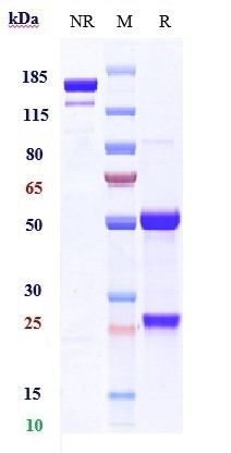
Recombinant Antibody
- 产品详情
- 实验流程
Application
| FC, Kinetics, Animal Model |
|---|---|
| Primary Accession | Q10589 |
| Reactivity | Human |
| Clonality | Monoclonal |
| Isotype | IgG1 |
| Calculated MW | 19769 Da |
| Target/Specificity | BST2 / CD317 |
|---|---|
| Endotoxin | < 0.001EU/ µg,determined by LAL method. |
| Conjugation | Unconjugated |
| Expression system | CHO Cell |
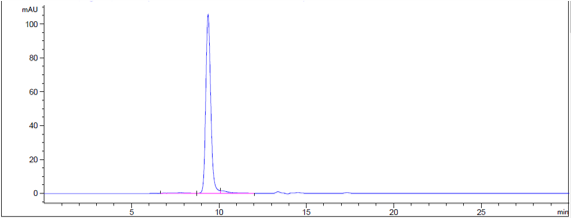
| Format | Purified monoclonal antibody supplied in 100mM Pro-Ac, 20mM Arg, pH5.0, without preservative.This antibody is purified through a protein A column. |
| Name | BST2 |
|---|---|
| Function | IFN-induced antiviral host restriction factor which efficiently blocks the release of diverse mammalian enveloped viruses by directly tethering nascent virions to the membranes of infected cells. Acts as a direct physical tether, holding virions to the cell membrane and linking virions to each other. The tethered virions can be internalized by endocytosis and subsequently degraded or they can remain on the cell surface. In either case, their spread as cell-free virions is restricted (PubMed:18200009, PubMed:18342597, PubMed:19036818, PubMed:19879838, PubMed:20019814, PubMed:20399176, PubMed:20419159, PubMed:20940320, PubMed:21529378, PubMed:22520941, PubMed:37922253). Its target viruses belong to diverse families, including retroviridae: human immunodeficiency virus type 1 (HIV-1), human immunodeficiency virus type 2 (HIV-2), simian immunodeficiency viruses (SIVs), equine infectious anemia virus (EIAV), feline immunodeficiency virus (FIV), prototype foamy virus (PFV), Mason-Pfizer monkey virus (MPMV), human T-cell leukemia virus type 1 (HTLV-1), Rous sarcoma virus (RSV) and murine leukemia virus (MLV), flavivirideae: hepatitis C virus (HCV), filoviridae: ebola virus (EBOV) and marburg virus (MARV), arenaviridae: lassa virus (LASV) and machupo virus (MACV), herpesviridae: kaposis sarcoma-associated herpesvirus (KSHV), rhabdoviridae: vesicular stomatitis virus (VSV), orthomyxoviridae: influenza A virus, paramyxoviridae: nipah virus, and coronaviridae: SARS-CoV (PubMed:18200009, PubMed:18342597, PubMed:19179289, PubMed:19879838, PubMed:20399176, PubMed:20419159, PubMed:20686043, PubMed:20943977, PubMed:21529378, PubMed:21621240, PubMed:22520941, PubMed:26378163, PubMed:31199522). Can inhibit cell surface proteolytic activity of MMP14 causing decreased activation of MMP15 which results in inhibition of cell growth and migration (PubMed:22065321). Can stimulate signaling by LILRA4/ILT7 and consequently provide negative feedback to the production of IFN by plasmacytoid dendritic cells in response to viral infection (PubMed:19564354, PubMed:26172439). Plays a role in the organization of the subapical actin cytoskeleton in polarized epithelial cells. Isoform 1 and isoform 2 are both effective viral restriction factors but have differing antiviral and signaling activities (PubMed:23028328, PubMed:26172439). Isoform 2 is resistant to HIV-1 Vpu-mediated degradation and restricts HIV-1 viral budding in the presence of Vpu (PubMed:23028328, PubMed:26172439). Isoform 1 acts as an activator of NF-kappa-B and this activity is inhibited by isoform 2 (PubMed:23028328). |
| Cellular Location | Golgi apparatus, trans-Golgi network. Cell membrane; Single-pass type II membrane protein. Cell membrane; Lipid- anchor, GPI-anchor. Membrane raft. Cytoplasm. Apical cell membrane. Note=Shuttles between the cell membrane, where it is present predominantly in membrane/lipid rafts, and the trans- Golgi network. Forms a complex with MMP14 and localizes to the cytoplasm |
| Tissue Location | Predominantly expressed in liver, lung, heart and placenta. Lower levels in pancreas, kidney, skeletal muscle and brain Overexpressed in multiple myeloma cells. Highly expressed during B-cell development, from pro-B precursors to plasma cells. Highly expressed on T-cells, monocytes, NK cells and dendritic cells (at protein level) |
Research Areas
For Research Use Only. Not For Use In Diagnostic Procedures.
Application Protocols
Provided below are standard protocols that you may find useful for product applications.
终于等到您。ABCEPTA(百远生物)抗体产品。
点击下方“我要评价 ”按钮提交您的反馈信息,您的反馈和评价是我们最宝贵的财富之一,
我们将在1-3个工作日内处理您的反馈信息。
如有疑问,联系:0512-88856768 tech-china@abcepta.com.
¥ 1,500.00
Cat# APR10282


