


 癌症的基本特征包括细胞增殖、血管生成、迁移、凋亡逃避机制和细胞永生等。找到癌症发生过程中这些通路的关键标记物和对应的抗体用于检测至关重要。
 为您推荐一个泛素化位点预测神器——泛素化分析工具,可以为您的蛋白的泛素化位点作出预测和评分。
 细胞自噬受体图形绘图工具为你的蛋白的细胞受体结合位点作出预测和评分,识别结合到自噬通路中的蛋白是非常重要的,便于让我们理解自噬在正常生理、病理过程中的作用,如发育、细胞分化、神经退化性疾病、压力条件下、感染和癌症。
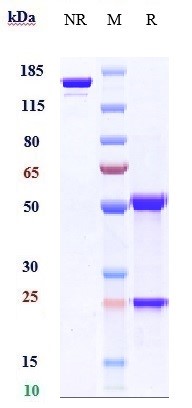
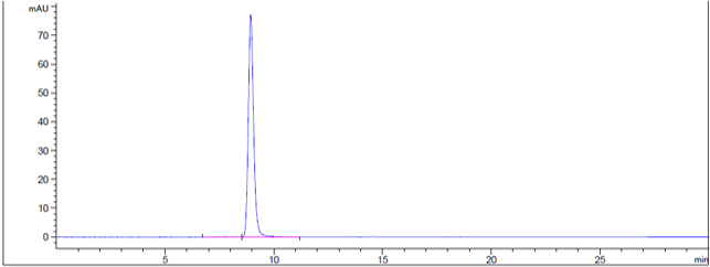
Anti-PMEL Reference Antibody (Genentech anti-PMEL17)
Recombinant Antibody
- 产品详情
- 实验流程
Application
| FC, Kinetics, Animal Model |
|---|---|
| Primary Accession | P40967 |
| Reactivity | Human, Mouse, Rat |
| Clonality | Monoclonal |
| Isotype | IgG1 |
| Calculated MW | 70255 Da |
| Target/Specificity | PMEL |
|---|---|
| Endotoxin | < 0.001EU/ µg,determined by LAL method. |
| Conjugation | Unconjugated |
| Expression system | CHO Cell |
| Format | Purified monoclonal antibody supplied in PBS, pH6.0, without preservative.This antibody is purified through a protein A column. |
| Name | PMEL |
|---|---|
| Synonyms | D12S53E, PMEL17, SILV |
| Function | Forms physiological amyloids that play a central role in melanosome morphogenesis and pigmentation. The maturation of unpigmented premelanosomes from stage I to II is marked by assembly of processed amyloidogenic fragments into parallel fibrillar sheets, which elongate the vesicle into a striated ellipsoidal shape. In pigmented stage III and IV melanosomes, the amyloid matrix serves as a platform where eumelanin precursors accumulate at high local concentrations for pigment formation. May prevent pigmentation-associated toxicity by sequestering toxic reaction intermediates of eumelanin biosynthesis pathway. |
| Cellular Location | Endoplasmic reticulum membrane; Single-pass type I membrane protein. Golgi apparatus, cis-Golgi network membrane; Single-pass type I membrane protein. Endosome, multivesicular body. Melanosome Extracellular vesicle. Secreted. Note=Identified by mass spectrometry in melanosome fractions from stage I to stage IV (PubMed:17081065) Localizes predominantly to intralumenal vesicles (ILVs) within multivesicular bodies. Associates with ILVs found within the lumen of premelanosomes and melanosomes and particularly in compartments that serve as precursors to the striated stage II premelanosomes (PubMed:11694580, PubMed:12643545). Sorted to stage I melanosomes following its processing in the ER and cis-Golgi (PubMed:15096515) Transiently expressed at the cell surface before targeting to early melanosomes (PubMed:16760433, PubMed:30988362). Colocalizes with BACE2 in stage I and II melanosomes (PubMed:23754390). Colocalizes with CD63 and APOE at exosomes and in intraluminal vesicles within multivesicular endosomes (PubMed:21962903, PubMed:26387950) |
| Tissue Location | Normally expressed at low levels in quiescent adult melanocytes but overexpressed by proliferating neonatal melanocytes and during tumor growth. Overexpressed in melanomas. Some expression was found in dysplastic nevi. |
Research Areas
For Research Use Only. Not For Use In Diagnostic Procedures.
Application Protocols
Provided below are standard protocols that you may find useful for product applications.
终于等到您。ABCEPTA(百远生物)抗体产品。
点击下方“我要评价 ”按钮提交您的反馈信息,您的反馈和评价是我们最宝贵的财富之一,
我们将在1-3个工作日内处理您的反馈信息。
如有疑问,联系:0512-88856768 tech-china@abcepta.com.
¥ 1,500.00
Cat# APR11014


