


 癌症的基本特征包括细胞增殖、血管生成、迁移、凋亡逃避机制和细胞永生等。找到癌症发生过程中这些通路的关键标记物和对应的抗体用于检测至关重要。
 为您推荐一个泛素化位点预测神器——泛素化分析工具,可以为您的蛋白的泛素化位点作出预测和评分。
 细胞自噬受体图形绘图工具为你的蛋白的细胞受体结合位点作出预测和评分,识别结合到自噬通路中的蛋白是非常重要的,便于让我们理解自噬在正常生理、病理过程中的作用,如发育、细胞分化、神经退化性疾病、压力条件下、感染和癌症。
> 首页 > 产品 > 一抗 > 精选抗体 > Anti-TNFRSF21 / DR6 / CD358 Reference Antibody (Abbvie patent anti-TNFRSF21)
Anti-TNFRSF21 / DR6 / CD358 Reference Antibody (Abbvie patent anti-TNFRSF21)
Recombinant Antibody
- 产品详情
- 实验流程
Application
| FC, Kinetics, Animal Model |
|---|---|
| Primary Accession | O75509 |
| Reactivity | Human |
| Clonality | Monoclonal |
| Isotype | IgG1 |
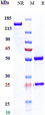
| Calculated MW | 71845 Da |
| Target/Specificity | TNFRSF21 / DR6 / CD358 |
|---|---|
| Endotoxin | < 0.001EU/ µg,determined by LAL method. |
| Conjugation | Unconjugated |
| Expression system | CHO Cell |
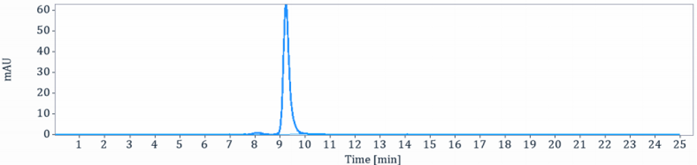
| Format | Purified monoclonal antibody supplied in PBS, pH6.0, without preservative.This antibody is purified through a protein A column. |
| Name | TNFRSF21 |
|---|---|
| Synonyms | DR6 |
| Function | Promotes apoptosis, possibly via a pathway that involves the activation of NF-kappa-B. Can also promote apoptosis mediated by BAX and by the release of cytochrome c from the mitochondria into the cytoplasm. Trophic-factor deprivation triggers the cleavage of surface APP by beta-secretase to release sAPP-beta which is further cleaved to release an N-terminal fragment of APP (N-APP). Negatively regulates oligodendrocyte survival, maturation and myelination. Plays a role in signaling cascades triggered by stimulation of T-cell receptors, in the adaptive immune response and in the regulation of T-cell differentiation and proliferation. Negatively regulates T-cell responses and the release of cytokines such as IL4, IL5, IL10, IL13 and IFNG by Th2 cells. Negatively regulates the production of IgG, IgM and IgM in response to antigens. May inhibit the activation of JNK in response to T-cell stimulation. Also acts as a regulator of pyroptosis: recruits CASP8 in response to reactive oxygen species (ROS) and subsequent oxidation, leading to activation of GSDMC (PubMed:34012073). |
| Cellular Location | Cell membrane; Single-pass type I membrane protein Note=Endocytosed following oxidation in response to reactive oxygen species (ROS). |
| Tissue Location | Detected in fetal spinal cord and in brain neurons, with higher levels in brain from Alzheimer disease patients (at protein level). Highly expressed in heart, brain, placenta, pancreas, lymph node, thymus and prostate. Detected at lower levels in lung, skeletal muscle, kidney, testis, uterus, small intestine, colon, spleen, bone marrow and fetal liver. Very low levels were found in adult liver and peripheral blood leukocytes. |
Research Areas
For Research Use Only. Not For Use In Diagnostic Procedures.
Application Protocols
Provided below are standard protocols that you may find useful for product applications.
终于等到您。ABCEPTA(百远生物)抗体产品。
点击下方“我要评价 ”按钮提交您的反馈信息,您的反馈和评价是我们最宝贵的财富之一,
我们将在1-3个工作日内处理您的反馈信息。
如有疑问,联系:0512-88856768 tech-china@abcepta.com.
¥ 1,500.00
Cat# APR11054


