


 癌症的基本特征包括细胞增殖、血管生成、迁移、凋亡逃避机制和细胞永生等。找到癌症发生过程中这些通路的关键标记物和对应的抗体用于检测至关重要。
 为您推荐一个泛素化位点预测神器——泛素化分析工具,可以为您的蛋白的泛素化位点作出预测和评分。
 细胞自噬受体图形绘图工具为你的蛋白的细胞受体结合位点作出预测和评分,识别结合到自噬通路中的蛋白是非常重要的,便于让我们理解自噬在正常生理、病理过程中的作用,如发育、细胞分化、神经退化性疾病、压力条件下、感染和癌症。
Anti-CNTFR Reference Antibody (Regeneron patent anti-CNTFR)
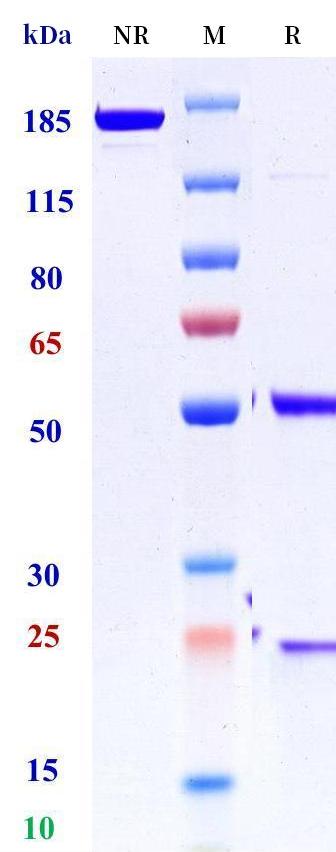
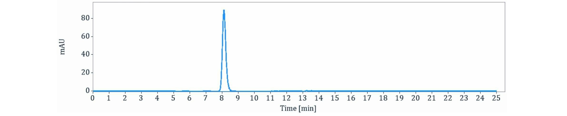
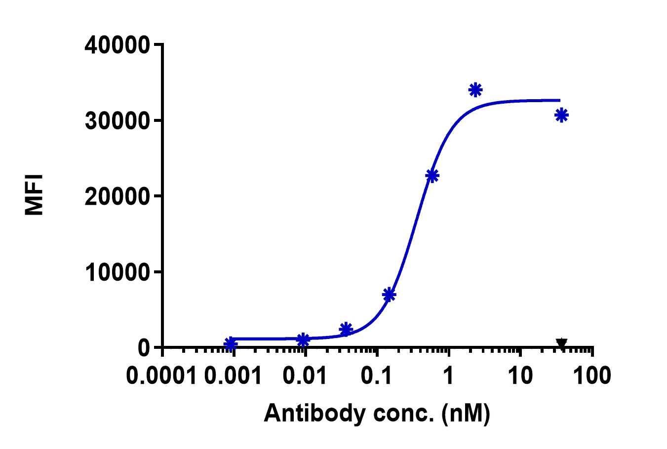
- 产品详情
- 实验流程
Application 
| Functional assay, Kinetics (SPR), Kinetics (BLI), FACS, E, FTA |
|---|---|
| Primary Accession | P26992 |
| Reactivity | Human |
| Clonality | Monoclonal |
| Isotype | IgG4SP |
| Calculated MW | 40633 Da |
| Target/Specificity | CNTFR |
|---|---|
| Expression system | CHO |
For Research Use Only. Not For Use In Diagnostic Procedures.
Patent Notice: The variable region sequence of this antibody is derived from a published third-party patent. This product is manufactured independently as a Research Use Only (RUO) reagent and is not licensed, sponsored, or endorsed by the patent holder. It is intended solely for research purposes. Customers considering any commercial, diagnostic, or therapeutic use should independently evaluate freedom-to-operate with respect to relevant third-party patents.
| Name | CNTFR {ECO:0000303|PubMed:7774913, ECO:0000312|EMBL:AAA35707.1} |
|---|---|
| Function | Functions either as a membrane-anchored non-signaling receptor via a GPI anchor or, after proteolytic release, as a soluble ligand-binding chaperone, and mediates signaling of CNTF-related cytokines, including ciliary neurotrophic factor (CNTF), cardiotrophin- like cytokine factor 1 (CLCF1), and the heterodimeric cytokine formed by CLCF1 and the cytokine receptor-like factor 1 (CRLF1) (PubMed:10966616, PubMed:11285233, PubMed:11294841, PubMed:26858303, PubMed:31932351, PubMed:36930708, PubMed:7681218). Involved namely in cell survival of motor neurons and regulation of oligodendrocyte progenitor proliferation (PubMed:11285233, PubMed:19386761, PubMed:31932351). Functions within the ciliary neurotrophic factor receptor (CNTFR) complex as a membrane-anchored non-signaling receptor subunit associated with two other signaling receptor subunits IL6ST/gp130 and LIFR (PubMed:10966616, PubMed:11285233, PubMed:11294841, PubMed:26858303, PubMed:31932351, PubMed:36930708, PubMed:7681218). Also, functions within the humanin receptor complex as a membrane-anchored non-signaling receptor subunit associated with IL6ST/GP130 and IL27RA/WSX1 that mediates the MT-RNR2/humanin signaling which induces neuroprotection (PubMed:19386761). Alternatively functions as a soluble ligand-binding chaperone that binds ligand in the extracellular space, forming a cytokine complex which is delivered to the IL6ST/gp130 and LIFR signaling receptor subunits (PubMed:11285233, PubMed:31932351, PubMed:7681218). Mechanistically, ligand binding to the CNTFR induces dimerization of the IL6ST/gp130 and LIFR (or IL27RA/WSX1 in the case of MT-RNR2/humanin), which activate JAK tyrosine kinases (JAK1 or JAK2 and to lesser extent TYK2) bound to their intracellular domains (PubMed:10966616, PubMed:11285233, PubMed:11294841, PubMed:19386761, PubMed:7681218). These kinases subsequently phosphorylate IL6ST/gp130 and LIFR (or IL27RA/WSX1) (PubMed:10966616, PubMed:11285233, PubMed:11294841, PubMed:7681218). The tyrosine phosphorylated signaling receptors serve in turn as docking sites for recruitment and activation of signal transducer and activators of transcription (STAT3 and to lesser extent STAT1) (PubMed:10966616, PubMed:11285233, PubMed:11294841, PubMed:19386761, PubMed:7681218). |
| Cellular Location | Cell membrane; Lipid-anchor, GPI-anchor. Secreted. Note=Localized at cell membrane when associated with SORL1 (PubMed:26858303). Released from plasma membrane and extracellular vesicles (EV) by GDPD2 (PubMed:31932351) |
| Tissue Location | Nervous system and skeletal muscle (PubMed:1648265). Soluble form is detected in cerebrospinal fluid (CSF) and is released from skeletal muscle in response to peripheral nerve injury (PubMed:7681218). |
Provided below are standard protocols that you may find useful for product applications.

终于等到您。ABCEPTA(百远生物)抗体产品。
点击下方“我要评价 ”按钮提交您的反馈信息,您的反馈和评价是我们最宝贵的财富之一,
我们将在1-3个工作日内处理您的反馈信息。
如有疑问,联系:0512-88856768 tech-china@abcepta.com.


