


 癌症的基本特征包括细胞增殖、血管生成、迁移、凋亡逃避机制和细胞永生等。找到癌症发生过程中这些通路的关键标记物和对应的抗体用于检测至关重要。
 为您推荐一个泛素化位点预测神器——泛素化分析工具,可以为您的蛋白的泛素化位点作出预测和评分。
 细胞自噬受体图形绘图工具为你的蛋白的细胞受体结合位点作出预测和评分,识别结合到自噬通路中的蛋白是非常重要的,便于让我们理解自噬在正常生理、病理过程中的作用,如发育、细胞分化、神经退化性疾病、压力条件下、感染和癌症。
DR3 Antibody
- 产品详情
- 实验流程
- 背景知识
Application
| WB, IF, ICC, E |
|---|---|
| Primary Accession | Q93038 |
| Other Accession | AAQ88676, 37181738 |
| Reactivity | Human, Mouse |
| Host | Rabbit |
| Clonality | Polyclonal |
| Isotype | IgG |
| Calculated MW | 45385 Da |
| Conjugate | Unconjugated |
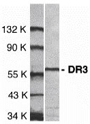
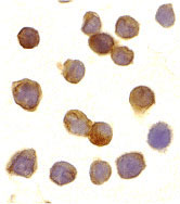
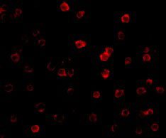
| Application Notes | DR3 antibody can be used for detection of DR3 expression by Western blot at 1 µg/mL. 59 kDa band should be detected. Antibody can also be used for immunocytochemistry starting at 10 µg/mL. For immunofluorescence start at 20 µg/mL. |
| Gene ID | 8718 |
|---|---|
| Other Names | DR3 Antibody: DR3, TR3, DDR3, LARD, APO-3, TRAMP, WSL-1, WSL-LR, TNFRSF12, APO3, DR3, WSL, WSL1, UNQ455/PRO779, Tumor necrosis factor receptor superfamily member 25, Apo-3, tumor necrosis factor receptor superfamily, member 25 |
| Target/Specificity | TNFRSF25; |
| Reconstitution & Storage | DR3 antibody can be stored at 4℃ for three months and -20℃, stable for up to one year. As with all antibodies care should be taken to avoid repeated freeze thaw cycles. Antibodies should not be exposed to prolonged high temperatures. |
| Precautions | DR3 Antibody is for research use only and not for use in diagnostic or therapeutic procedures. |
| Name | TNFRSF25 |
|---|---|
| Synonyms | APO3, DDR3, DR3, TNFRSF12, WSL, WSL1 |
| Function | Receptor for TNFSF12/APO3L/TWEAK. Interacts directly with the adapter TRADD. Mediates activation of NF-kappa-B and induces apoptosis. May play a role in regulating lymphocyte homeostasis. |
| Cellular Location | [Isoform 1]: Cell membrane; Single-pass type I membrane protein [Isoform 9]: Cell membrane; Single-pass type I membrane protein [Isoform 3]: Secreted. [Isoform 5]: Secreted. [Isoform 7]: Secreted. [Isoform 10]: Secreted. |
| Tissue Location | Abundantly expressed in thymocytes and lymphocytes. Detected in lymphocyte-rich tissues such as thymus, colon, intestine, and spleen. Also found in the prostate |
For Research Use Only. Not For Use In Diagnostic Procedures.
Provided below are standard protocols that you may find useful for product applications.
BACKGROUND
DR3 Antibody: Apoptosis, or programmed cell death, occurs during normal cellular differentiation and development of multicellular organisms. Apoptosis is induced by certain cytokines including TNF and Fas ligand of the TNF family through their death domain containing receptors, TNFR1 and Fas. A novel cell death receptor was recently identified by several groups independently and designated DR3, Wsl-1, Apo-3, TRAMP and LARD1-5. The ligand for this novel death receptor has been defined as TWEAK, also termed Apo3L. DR3 is highly expressed in the tissues enriched in lymphocytes including PBL, thymus and spleen. Like TNFR1, DR3 induces apoptosis and NF-κB activation.
REFERENCES
Chinnaiyan AM; O'Rourke K; Yu GL; Lyons RH; Garg M; Duan DR; Xing L; Gentz R; Ni J; Dixit VM. Science, 1996;274:990-2.
Kitson J; Raven T; Jiang YP; Goeddel DV; Giles KM; Pun KT; Grinham CJ; Brown R; Farrow SN. Nature, 1996;384:372-5.
Marsters SA; Sheridan JP; Donahue CJ; Pitti RM; Gray CL; Goddard AD; Bauer KD; Ashkenazi A. Curr Biol, 1996;6:1669-76.
Bodmer JL; Burns K; Schneider P; Hofmann K; Steiner V; Thome M; Bornand T; Hahne M; Schroter M; Becker K; et al. Immunity, 1997;6:79-88.
终于等到您。ABCEPTA(百远生物)抗体产品。
点击下方“我要评价 ”按钮提交您的反馈信息,您的反馈和评价是我们最宝贵的财富之一,
我们将在1-3个工作日内处理您的反馈信息。
如有疑问,联系:0512-88856768 tech-china@abcepta.com.


