


 癌症的基本特征包括细胞增殖、血管生成、迁移、凋亡逃避机制和细胞永生等。找到癌症发生过程中这些通路的关键标记物和对应的抗体用于检测至关重要。
 为您推荐一个泛素化位点预测神器——泛素化分析工具,可以为您的蛋白的泛素化位点作出预测和评分。
 细胞自噬受体图形绘图工具为你的蛋白的细胞受体结合位点作出预测和评分,识别结合到自噬通路中的蛋白是非常重要的,便于让我们理解自噬在正常生理、病理过程中的作用,如发育、细胞分化、神经退化性疾病、压力条件下、感染和癌症。
IKK alpha Antibody
- 产品详情
- 实验流程
- 背景知识
Application
| WB, IF, ICC, E |
|---|---|
| Primary Accession | O15111 |
| Other Accession | AF009225, 2327068 |
| Reactivity | Human |
| Host | Rabbit |
| Clonality | Polyclonal |
| Isotype | IgG |
| Calculated MW | 84640 Da |
| Conjugate | Unconjugated |
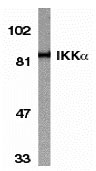
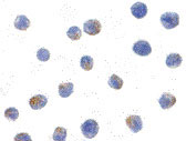
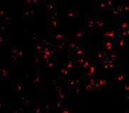
| Application Notes | IKK alpha can be used for detection of IKK alpha by Western blot at 1 µg/mL. An 85 kDa band should be detected. Antibody can also be used for immunocytochemistry starting at 1 µg/mL. For immunofluorescence start at 20 µg/mL. |
| Gene ID | 1147 |
|---|---|
| Other Names | IKK alpha Antibody: IKK1, IKKA, IKBKA, TCF16, NFKBIKA, IKK-alpha, Inhibitor of nuclear factor kappa-B kinase subunit alpha, Conserved helix-loop-helix ubiquitous kinase, I-kappa-B kinase alpha, conserved helix-loop-helix ubiquitous kinase |
| Target/Specificity | CHUK; Antibody has no cross response to IKKb or IKKg. |
| Reconstitution & Storage | IKK alpha antibody can be stored at 4℃ for three months and -20℃, stable for up to one year. As with all antibodies care should be taken to avoid repeated freeze thaw cycles. Antibodies should not be exposed to prolonged high temperatures. |
| Precautions | IKK alpha Antibody is for research use only and not for use in diagnostic or therapeutic procedures. |
| Name | CHUK |
|---|---|
| Synonyms | IKKA, TCF16 |
| Function | Serine kinase that plays an essential role in the NF-kappa-B signaling pathway which is activated by multiple stimuli such as inflammatory cytokines, bacterial or viral products, DNA damages or other cellular stresses (PubMed:18626576, PubMed:9244310, PubMed:9252186, PubMed:9346484). Acts as a part of the canonical IKK complex in the conventional pathway of NF-kappa-B activation and phosphorylates inhibitors of NF-kappa-B on serine residues (PubMed:18626576, PubMed:35952808, PubMed:9244310, PubMed:9252186, PubMed:9346484). These modifications allow polyubiquitination of the inhibitors and subsequent degradation by the proteasome (PubMed:18626576, PubMed:9244310, PubMed:9252186, PubMed:9346484). In turn, free NF-kappa-B is translocated into the nucleus and activates the transcription of hundreds of genes involved in immune response, growth control, or protection against apoptosis (PubMed:18626576, PubMed:9244310, PubMed:9252186, PubMed:9346484). Negatively regulates the pathway by phosphorylating the scaffold protein TAXBP1 and thus promoting the assembly of the A20/TNFAIP3 ubiquitin-editing complex (composed of A20/TNFAIP3, TAX1BP1, and the E3 ligases ITCH and RNF11) (PubMed:21765415). Therefore, CHUK plays a key role in the negative feedback of NF-kappa-B canonical signaling to limit inflammatory gene activation. As part of the non-canonical pathway of NF-kappa-B activation, the MAP3K14-activated CHUK/IKKA homodimer phosphorylates NFKB2/p100 associated with RelB, inducing its proteolytic processing to NFKB2/p52 and the formation of NF-kappa-B RelB-p52 complexes (PubMed:20501937). In turn, these complexes regulate genes encoding molecules involved in B-cell survival and lymphoid organogenesis. Also participates in the negative feedback of the non-canonical NF-kappa-B signaling pathway by phosphorylating and destabilizing MAP3K14/NIK. Within the nucleus, phosphorylates CREBBP and consequently increases both its transcriptional and histone acetyltransferase activities (PubMed:17434128). Modulates chromatin accessibility at NF-kappa-B- responsive promoters by phosphorylating histones H3 at 'Ser-10' that are subsequently acetylated at 'Lys-14' by CREBBP (PubMed:12789342). Additionally, phosphorylates the CREBBP-interacting protein NCOA3. Also phosphorylates FOXO3 and may regulate this pro-apoptotic transcription factor (PubMed:15084260). Phosphorylates RIPK1 at 'Ser-25' which represses its kinase activity and consequently prevents TNF-mediated RIPK1-dependent cell death (By similarity). Phosphorylates AMBRA1 following mitophagy induction, promoting AMBRA1 interaction with ATG8 family proteins and its mitophagic activity (PubMed:30217973). |
| Cellular Location | Cytoplasm. Nucleus Note=Shuttles between the cytoplasm and the nucleus |
| Tissue Location | Widely expressed. |
For Research Use Only. Not For Use In Diagnostic Procedures.
Provided below are standard protocols that you may find useful for product applications.
BACKGROUND
IKK alpha Antibody: Nuclear factor kappa B (NF-κB) is a ubiquitous transcription factor and an essential mediator of gene expression during activation of immune and inflammatory responses. NF-κB mediates the expression of a great variety of genes in response to extracellular stimuli including IL-1, TNFa, and bacteria product LPS. NF-κB is associated with IκB proteins in the cell cytoplasm, which inhibit NF-κB activity. The long-sought IκB kinase (IKK), which phosphorylates IκB, and mediates IκB degradation and NF-κB activation, was recently identified by several laboratories. IKK is a serine protein kinase, and the IKK complex contains alpha and beta subunits (IKKα and IKKβ). IKKα and IKKβ interact with each other and both are essential for the NF-κB activation. IKKα specifically phosphorylates IkB-alpha. IKKα is expressed in variety of human tissues.
REFERENCES
DiDonato JA, Hayakawa M, Rothwarf DM, Zandi E, Karin M. A cytokine-responsive IκB kinase that activates the transcription factor NF-κB. Nature 1997;388:548-54
Regnier CH, Song HY, Gao X, Goeddel DV, Cao Z, Rothe M. Identification and characterization of an IκB kinase. Cell 1997;90:373-83
Zandi E, Rothwarf DM, Delhase M, Hayakawa M, Karin M. The IκB kinase complex (IKK) contains two kinase subunits, IKKα and IKKβ, necessary for IκB phosphorylation and NF-κB activation. Cell 1997;91:243-52
Woronicz JD, Gao X, Cao Z, Rothe M, Goeddel DY. IκB kinase-β: NF-κB activation and complex formation with IκB kinase-α and NIK. Science 1997;278:866-9
终于等到您。ABCEPTA(百远生物)抗体产品。
点击下方“我要评价 ”按钮提交您的反馈信息,您的反馈和评价是我们最宝贵的财富之一,
我们将在1-3个工作日内处理您的反馈信息。
如有疑问,联系:0512-88856768 tech-china@abcepta.com.


