


 癌症的基本特征包括细胞增殖、血管生成、迁移、凋亡逃避机制和细胞永生等。找到癌症发生过程中这些通路的关键标记物和对应的抗体用于检测至关重要。
 为您推荐一个泛素化位点预测神器——泛素化分析工具,可以为您的蛋白的泛素化位点作出预测和评分。
 细胞自噬受体图形绘图工具为你的蛋白的细胞受体结合位点作出预测和评分,识别结合到自噬通路中的蛋白是非常重要的,便于让我们理解自噬在正常生理、病理过程中的作用,如发育、细胞分化、神经退化性疾病、压力条件下、感染和癌症。
ZIPK Antibody
- 产品详情
- 实验流程
- 背景知识
Application
| WB, IF, ICC, E |
|---|---|
| Primary Accession | O43293 |
| Other Accession | BAA81746, 5162884 |
| Reactivity | Human, Mouse, Rat |
| Host | Rabbit |
| Clonality | Polyclonal |
| Isotype | IgG |
| Calculated MW | 52536 Da |
| Conjugate | Unconjugated |
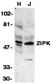
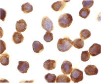
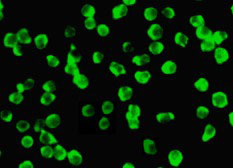
| Application Notes | ZIPK antibody can be used for detection of DNase II expression by Western blot at 1 µg/mL. An approximate 40 kDa band can be detected, which represents the pro-enzyme of DNase II. Antibody can also be used for immunocytochemistry starting at 10 µg/mL. For immunofluorescence start at 10 µg/mL. |
| Gene ID | 1613 |
|---|---|
| Other Names | ZIPK Antibody: ZIP, ZIPK, Death-associated protein kinase 3, DAP-like kinase, DAP kinase 3, death-associated protein kinase 3 |
| Target/Specificity | DAPK3; |
| Reconstitution & Storage | ZIPK antibody can be stored at 4℃ for three months and -20℃, stable for up to one year. As with all antibodies care should be taken to avoid repeated freeze thaw cycles. Antibodies should not be exposed to prolonged high temperatures. |
| Precautions | ZIPK Antibody is for research use only and not for use in diagnostic or therapeutic procedures. |
| Name | DAPK3 |
|---|---|
| Synonyms | ZIPK |
| Function | Serine/threonine kinase which is involved in the regulation of apoptosis, autophagy, transcription, translation and actin cytoskeleton reorganization. Involved in the regulation of smooth muscle contraction. Regulates both type I (caspase-dependent) apoptotic and type II (caspase-independent) autophagic cell deaths signal, depending on the cellular setting. Involved in regulation of starvation-induced autophagy. Regulates myosin phosphorylation in both smooth muscle and non-muscle cells. In smooth muscle, regulates myosin either directly by phosphorylating MYL12B and MYL9 or through inhibition of smooth muscle myosin phosphatase (SMPP1M) via phosphorylation of PPP1R12A; the inhibition of SMPP1M functions to enhance muscle responsiveness to Ca(2+) and promote a contractile state. Phosphorylates MYL12B in non-muscle cells leading to reorganization of actin cytoskeleton. Isoform 2 can phosphorylate myosin, PPP1R12A and MYL12B. Overexpression leads to condensation of actin stress fibers into thick bundles. Involved in actin filament focal adhesion dynamics. The function in both reorganization of actin cytoskeleton and focal adhesion dissolution is modulated by RhoD. Positively regulates canonical Wnt/beta-catenin signaling through interaction with NLK and TCF7L2. Phosphorylates RPL13A on 'Ser-77' upon interferon-gamma activation which is causing RPL13A release from the ribosome, RPL13A association with the GAIT complex and its subsequent involvement in transcript-selective translation inhibition. Enhances transcription from AR-responsive promoters in a hormone- and kinase- dependent manner. Involved in regulation of cell cycle progression and cell proliferation. May be a tumor suppressor. |
| Cellular Location | Nucleus. Nucleus, PML body {ECO:0000250|UniProtKB:O54784}. Cytoplasm, cytoskeleton, microtubule organizing center, centrosome {ECO:0000250|UniProtKB:O54784}. Chromosome, centromere. Cytoplasm. Cytoplasm, cytoskeleton, spindle. Midbody Note=Predominantly localizes to the cytoplasm but can shuttle between the nucleus and cytoplasm; cytoplasmic localization is promoted by phosphorylation at Thr-299 and involves Rho/Rock signaling [Isoform 2]: Nucleus. Cytoplasm |
| Tissue Location | Widely expressed. Isoform 1 and isoform 2 are expressed in the bladder smooth muscle. |
For Research Use Only. Not For Use In Diagnostic Procedures.
Provided below are standard protocols that you may find useful for product applications.
BACKGROUND
ZIPK Antibody: Apoptosis is mediated by death domain containing adapter molecules and a caspase family of proteases. Certain serine/threonine protein kinases, such as ASK-1 and RIP, are mediators of apoptosis. A novel serine/threonine kinase that mediates apoptosis was recently identified and designated ZIP kinase. ZIP kinase contains an N-terminal kinase domain and a C-terminal leucine zipper structure and binds to ATF4 that is a member of ATF/CREB family. ZIP kinase has high sequence homology to DAP kinase (death-associated protein kinase), which is a mediator of apoptosis induced by gamma interferon. Overexpression of ZIP kinase induces apoptosis. ZIP and DAP kinases represent a novel kinase family, which mediates apoptosis through their catalytic activities. The messenger RNA was ubiquitously expressed in various tissues.
REFERENCES
Kawai T, Matsumoto M, Takeda K, Sanjo H, Akira S. ZIP kinase, a novel serine/threonine kinase which mediates apoptosis. Mol Cell Biol 1998;18:1642-51 (RD1299)
终于等到您。ABCEPTA(百远生物)抗体产品。
点击下方“我要评价 ”按钮提交您的反馈信息,您的反馈和评价是我们最宝贵的财富之一,
我们将在1-3个工作日内处理您的反馈信息。
如有疑问,联系:0512-88856768 tech-china@abcepta.com.


