


 癌症的基本特征包括细胞增殖、血管生成、迁移、凋亡逃避机制和细胞永生等。找到癌症发生过程中这些通路的关键标记物和对应的抗体用于检测至关重要。
 为您推荐一个泛素化位点预测神器——泛素化分析工具,可以为您的蛋白的泛素化位点作出预测和评分。
 细胞自噬受体图形绘图工具为你的蛋白的细胞受体结合位点作出预测和评分,识别结合到自噬通路中的蛋白是非常重要的,便于让我们理解自噬在正常生理、病理过程中的作用,如发育、细胞分化、神经退化性疾病、压力条件下、感染和癌症。
CX3CR1 Antibody
- 产品详情
- 实验流程
- 背景知识
Application
| WB, IF, E, IHC-P |
|---|---|
| Primary Accession | P49238 |
| Other Accession | NP_001328, 4503171 |
| Reactivity | Human |
| Host | Rabbit |
| Clonality | Polyclonal |
| Isotype | IgG |
| Calculated MW | 39 KDa |
| Concentration (mg/ml) | 1 mg/mL |
| Conjugate | Unconjugated |
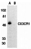
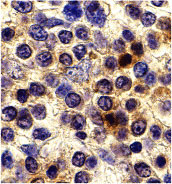
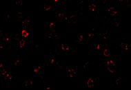
| Application Notes | CX3CR1 antibody can be used for detection of CX3CR1 by Western blot at 0.5 µg/mL. Antibody can also be used for immunohistochemistry starting at 10 µg/mL. For immunofluorescence start at 20 µg/mL. |
| Gene ID | 1524 |
|---|---|
| Other Names | CX3CR1 Antibody: V28, CCRL1, GPR13, CMKDR1, GPRV28, CMKBRL1, CX3C chemokine receptor 1, Beta chemokine receptor-like 1, C-X3-C CKR-1, chemokine (C-X3-C motif) receptor 1 |
| Target/Specificity | CX3CR1; At least two isoforms of CX3CR1 are known to exist. |
| Reconstitution & Storage | CX3CR1 antibody can be stored at 4℃ for three months and -20℃, stable for up to one year. As with all antibodies care should be taken to avoid repeated freeze thaw cycles. Antibodies should not be exposed to prolonged high temperatures. |
| Precautions | CX3CR1 Antibody is for research use only and not for use in diagnostic or therapeutic procedures. |
For Research Use Only. Not For Use In Diagnostic Procedures.
Provided below are standard protocols that you may find useful for product applications.
BACKGROUND
CX3CR1 Antibody: CX3CR1 is one of the chemokine receptors that are required as coreceptors for HIV infection. The genes encoding human, mouse, and rat CX3CR1 were cloned and designated V28 and CMKBRL1, CX3CR1, and RBS11, respectively. The encoded seven transmembrane protein was initially identified as the receptor for the transmembrane molecule fractalkine, and renamed CX3CR1. CX3CR1 is known to serve as a coreceptor for HIV-1 and HIV-2 envelope fusion and virus infection, which can be inhibited by fractokine. CX3CR1 mediates leukocyte migration and adhesion. CX3CR1 is expressed in a variety of human tissues and cell lines.
REFERENCES
Raport CJ, Schweickart VL, Eddy RL JR, et al. The orphan G-protein-coupled receptor-encoding gene V28 is closely related to genes for chemokine receptors and is expressed in lymphoid and neural tissues. Gene 1995; 163:295-9
Combadiere C, Ahuja SK , and Murphy PM. Cloning, chromosomal localization, and RNA expression of a human β chemokine receptor-like gene. DNA Cell Biol. 1995;14:673-80
Combadiere C, Gao J, Tiffany HL, et al. Gene cloning, RNA distribution, and functional expression of mCX3CR1, a mouse chemotactic receptor for the CX3C chemokine fractalkine. Biochem. Biophys. Res. Commun. 1998; 253:728-32
Harrison JK, Barber CM, and Lynch KR. cDNA cloning of a G-protein-coupled receptor expressed in rat spinal cord and brain related to chemokine receptors. Neurosci. Lett. 1994; 169:85-9
终于等到您。ABCEPTA(百远生物)抗体产品。
点击下方“我要评价 ”按钮提交您的反馈信息,您的反馈和评价是我们最宝贵的财富之一,
我们将在1-3个工作日内处理您的反馈信息。
如有疑问,联系:0512-88856768 tech-china@abcepta.com.


