


 癌症的基本特征包括细胞增殖、血管生成、迁移、凋亡逃避机制和细胞永生等。找到癌症发生过程中这些通路的关键标记物和对应的抗体用于检测至关重要。
 为您推荐一个泛素化位点预测神器——泛素化分析工具,可以为您的蛋白的泛素化位点作出预测和评分。
 细胞自噬受体图形绘图工具为你的蛋白的细胞受体结合位点作出预测和评分,识别结合到自噬通路中的蛋白是非常重要的,便于让我们理解自噬在正常生理、病理过程中的作用,如发育、细胞分化、神经退化性疾病、压力条件下、感染和癌症。
Toso Antibody
- 产品详情
- 实验流程
- 背景知识
Application
| WB, E |
|---|---|
| Primary Accession | O60667 |
| Other Accession | NP_005440, 4885641 |
| Reactivity | Human |
| Host | Rabbit |
| Clonality | Polyclonal |
| Isotype | IgG |
| Calculated MW | 43146 Da |
| Conjugate | Unconjugated |
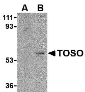
| Application Notes | Toso antibody can be used for detection of Toso by Western blot at 1 µg/mL. Despite its predicted molecular weight, Toso often migrates at 60 kDa in SDS-PAGE. |
| Gene ID | 9214 |
|---|---|
| Other Names | Toso Antibody: FCMR, TOSO, Fas apoptotic inhibitory molecule 3, Regulator of Fas-induced apoptosis Toso, Fas apoptotic inhibitory molecule 3 |
| Target/Specificity | FAIM3; |
| Reconstitution & Storage | Toso antibody can be stored at 4℃ for three months and -20℃, stable for up to one year. As with all antibodies care should be taken to avoid repeated freeze thaw cycles. Antibodies should not be exposed to prolonged high temperatures. |
| Precautions | Toso Antibody is for research use only and not for use in diagnostic or therapeutic procedures. |
| Name | FCMR {ECO:0000303|PubMed:25888699, ECO:0000312|HGNC:HGNC:14315} |
|---|---|
| Function | High-affinity Fc receptor for immunoglobulin M (IgM), both secreted and membrane-bound IgM (PubMed:19858324, PubMed:22675200, PubMed:36949194, PubMed:37095205). Primarily regulates IgM transport and homeostasis. In lymphoid cells, enables exocytosis of membrane- bound IgM on the plasma membrane as well as endocytosis of IgM-antigen complexes toward lysosomes for degradation. In mucosal epithelium, mediates retrotranscytosis of antigen-IgM complexes across mucosal M cells toward antigen-presenting cells in mucosal lymphoid tissues (PubMed:21908732, PubMed:28230186). Triggers costimulatory signaling and mediates most of IgM effector functions involved in B cell development and primary immune response to infection. Likely limits tonic IgM BCR signaling to self-antigens for proper negative selection of autoreactive B cells in the bone marrow and for the maintenance of regulatory B cell pool in peripheral lymphoid organs. Mediates antibody responses to T cell-dependent and T cell-independent antigens and promotes induction of an efficient neutralizing IgG response. Engages in cross-talk with antigen-receptor signaling via the non-canonical NF- kappa-B, MAP kinases and calcium signaling pathways (PubMed:19858324, PubMed:22675200, PubMed:25601920, PubMed:30840890). |
| Cellular Location | Cell membrane; Single-pass membrane protein. Early endosome membrane; Single-pass membrane protein. Golgi apparatus, trans- Golgi network membrane; Single-pass membrane protein. Lysosome membrane; Single-pass membrane protein. Note=Continuously recycles between cytoplasmic pool and the plasma membrane to bind as much IgM as possible |
| Tissue Location | Expressed by CD19-positive B cells and CD4-positive and CD8-positive T cell populations in primary and secondary lymphoid tissues (at protein level). Among B cell subsets, detected in a subset of bone marrow pro- and pre-B cells, in most follicular and memory B cells and in a small subset of germinal center B cells (at protein level). Expressed at lower levels in CD56-positive NK cells (at protein level) (PubMed:19858324, PubMed:21908732, PubMed:22675200, PubMed:30840890). Expressed in lymph nodes, lung, thymus and kidneys Very weak expression detected in spleen, liver, heart, and salivary gland. |
For Research Use Only. Not For Use In Diagnostic Procedures.
Provided below are standard protocols that you may find useful for product applications.
BACKGROUND
Toso Antibody: Apoptosis is an important process by which normal tissue homeostasis and function are maintained. One of the major signals that regulate this process is mediated by the activation of the Fas receptor by its ligand. This leads to the formation of a Fas-associated death domain (FADD)- containing death-inducing signaling complex and the activation of caspase-8, which in turn activates downstream effector caspases, such as caspase-3 and -7. Recent experiments have shown that overexpression of Toso, a novel regulator of Fas-induced apoptosis in lymphoid cells, in Jurkat cells as well as transgenic mice render these cells resistant to Fas-induced apoptosis but not to TRAIL-induced apoptosis. Furthermore, Toso was found to associate with FADD, suggesting that Toso functions by disrupting the formation of the death-inducing signaling complex.
REFERENCES
Curtin JF and Cotter TG. Live and let die: regulatory mechanisms in Fas-mediated apoptosis. Cell Signal. 2003; 15:983-92.
Hitoshi Y, Lorens J, Kitada S-I, et al. Toso, a cell surface, specific regulator of Fas-induced apoptosis in T cells. Immunity 1998; 8:461-71.
Song Y and Jacob CO. The mouse cell surface protein Toso regulates Fas/Fas ligand-induced apoptosis through its binding to Fas-associated death domain. J. Biol. Chem.2005; 280:9618-26.
终于等到您。ABCEPTA(百远生物)抗体产品。
点击下方“我要评价 ”按钮提交您的反馈信息,您的反馈和评价是我们最宝贵的财富之一,
我们将在1-3个工作日内处理您的反馈信息。
如有疑问,联系:0512-88856768 tech-china@abcepta.com.


