


 癌症的基本特征包括细胞增殖、血管生成、迁移、凋亡逃避机制和细胞永生等。找到癌症发生过程中这些通路的关键标记物和对应的抗体用于检测至关重要。
 为您推荐一个泛素化位点预测神器——泛素化分析工具,可以为您的蛋白的泛素化位点作出预测和评分。
 细胞自噬受体图形绘图工具为你的蛋白的细胞受体结合位点作出预测和评分,识别结合到自噬通路中的蛋白是非常重要的,便于让我们理解自噬在正常生理、病理过程中的作用,如发育、细胞分化、神经退化性疾病、压力条件下、感染和癌症。
F1A alpha Antibody
- 产品详情
- 实验流程
- 背景知识
Application
| WB, IF, E, IHC-P |
|---|---|
| Primary Accession | Q9UK73 |
| Other Accession | AAF05314, 6175869 |
| Reactivity | Human, Mouse, Rat |
| Host | Rabbit |
| Clonality | Polyclonal |
| Isotype | IgG |
| Calculated MW | 70264 Da |
| Conjugate | Unconjugated |
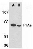
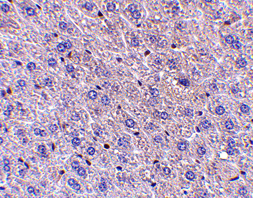
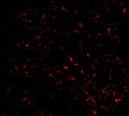
| Application Notes | F1Ab antibody can be used for detection of F1Ab by Western blot at 1 µg/mL. An approximately 70 kDa band can be detected. Antibody can also be used for immunohistochemistry starting at 5 µg/mL. For immunofluorescence start at 20 µg/mL. |
| Gene ID | 10116 |
|---|---|
| Other Names | F1A alpha Antibody: F1AA, F1A-ALPHA, FEM1-beta, F1AA, KIAA0396, Protein fem-1 homolog B, FEM1b, fem-1 homolog b (C. elegans) |
| Target/Specificity | FEM1B; |
| Reconstitution & Storage | F1A alpha antibody can be stored at 4℃ for three months and -20℃, stable for up to one year. As with all antibodies care should be taken to avoid repeated freeze thaw cycles. Antibodies should not be exposed to prolonged high temperatures. |
| Precautions | F1A alpha Antibody is for research use only and not for use in diagnostic or therapeutic procedures. |
| Name | FEM1B {ECO:0000303|PubMed:10623617, ECO:0000312|HGNC:HGNC:3649} |
|---|---|
| Function | Substrate-recognition component of a Cul2-RING (CRL2) E3 ubiquitin-protein ligase complex of the DesCEND (destruction via C-end degrons) pathway, which recognizes a C-degron located at the extreme C terminus of target proteins, leading to their ubiquitination and degradation (PubMed:29779948, PubMed:33398168, PubMed:33398170). The C- degron recognized by the DesCEND pathway is usually a motif of less than ten residues and can be present in full-length proteins, truncated proteins or proteolytically cleaved forms (PubMed:29779948, PubMed:33398168, PubMed:33398170). The CRL2(FEM1B) complex specifically recognizes proteins ending with -Gly-Leu-Asp-Arg, such as CDK5R1, leading to their ubiquitination and degradation (PubMed:33398168, PubMed:33398170). Also acts as a regulator of the reductive stress response by mediating ubiquitination of reduced FNIP1: in response to reductive stress, the CRL2(FEM1B) complex specifically recognizes a conserved Cys degron in FNIP1 when this degron is reduced, leading to FNIP1 degradation and subsequent activation of mitochondria to recalibrate reactive oxygen species (ROS) (By similarity). Mechanistically, recognizes and binds reduced FNIP1 through two interface zinc ions, which act as a molecular glue that recruit reduced FNIP1 to FEM1B (By similarity). Promotes ubiquitination of GLI1, suppressing GLI1 transcriptional activator activity (PubMed:24076122). Promotes ubiquitination and degradation of ANKRD37 (By similarity). Promotes ubiquitination and degradation of SLBP (PubMed:28118078). Involved in apoptosis by acting as a death receptor-associated protein that mediates apoptosis (PubMed:10542291). Also involved in glucose homeostasis in pancreatic islet (By similarity). May also act as an adapter/mediator in replication stress-induced signaling that leads to the activation of CHEK1 (PubMed:19330022). |
| Cellular Location | Cytoplasm. Nucleus Note=In the nucleus, the protein level increased slightly after camptothecin (CPT) treatment (PubMed:19330022). Associated with chromatin (PubMed:19330022). |
| Tissue Location | Widely expressed (PubMed:10542291). Highly expressed in testis (PubMed:10542291). Weakly expressed in other tissues (PubMed:10542291). |
For Research Use Only. Not For Use In Diagnostic Procedures.
Provided below are standard protocols that you may find useful for product applications.
BACKGROUND
F1A alpha Antibody: Fas and tumor necrosis factor receptor 1 (TNFR1) are two prototype members in the death receptor family. A novel protein that associates with the intracellular domains of Fas and TNFR1 was recently identified and designated F1Aalpha and FEM1β. F1Aalpha/FEM1β is the homologue of C. elegans sex determining protein FEM-1. FEM-1/F1Aalpha is cleaved by CED-3 and caspase. FEM-1/F1Aalpha associates with CED-4 and its mammalian homologue Apaf-1. Overexpression of F1Aalpha induces apoptosis. F1Aalpha is therefore a novel member of the death receptor associated protein that mediates apoptosis. F1Aalpha is expressed in a variety of human and mouse tissues.
REFERENCES
Chan SL, Tan KO, Zhang L, Yee KS , Ronca F, Chan MY, Yu VC. F1Aα, a death receptor-binding protein homologous to the Caenorhabditis elegans sex-determining protein, FEM-1, is a caspase substrate that mediates apoptosis. J Biol Chem. 1999;274(45):32461-8.
Ventura-Holman T, Maher JF. Sequence, organization, and expression of the human FEM1B gene. Biochem Biophys Res Commun 2000;267(1):317-20
Chan SL, Yee KS , Tan KM, Yu VC. The Caenorhabditis elegans sex determination protein FEM-1 is a CED-3 substrate that associates with CED-4 and mediates apoptosis in mammalian cells. J Biol Chem. 2000;275(24):17925-8. (WD0102)
终于等到您。ABCEPTA(百远生物)抗体产品。
点击下方“我要评价 ”按钮提交您的反馈信息,您的反馈和评价是我们最宝贵的财富之一,
我们将在1-3个工作日内处理您的反馈信息。
如有疑问,联系:0512-88856768 tech-china@abcepta.com.


