


 癌症的基本特征包括细胞增殖、血管生成、迁移、凋亡逃避机制和细胞永生等。找到癌症发生过程中这些通路的关键标记物和对应的抗体用于检测至关重要。
 为您推荐一个泛素化位点预测神器——泛素化分析工具,可以为您的蛋白的泛素化位点作出预测和评分。
 细胞自噬受体图形绘图工具为你的蛋白的细胞受体结合位点作出预测和评分,识别结合到自噬通路中的蛋白是非常重要的,便于让我们理解自噬在正常生理、病理过程中的作用,如发育、细胞分化、神经退化性疾病、压力条件下、感染和癌症。
PERP Antibody
- 产品详情
- 实验流程
- 背景知识
Application 
| WB, ICC, E |
|---|---|
| Primary Accession | Q9JK95 |
| Other Accession | NP_071404, 11528520 |
| Reactivity | Human |
| Host | Rabbit |
| Clonality | Polyclonal |
| Isotype | IgG |
| Calculated MW | 21567 Da |
| Concentration (mg/ml) | 1 mg/mL |
| Conjugate | Unconjugated |
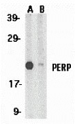
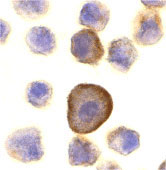
| Application Notes | PERP antibody can be used for detection of PERP by Western blot at 1 µg/mL. Antibody can also be used for immunocytochemistry starting at 10 µg/mL. |
| Gene ID | 64058 |
|---|---|
| Other Names | PERP Antibody: THW, KCP1, PIGPC1, KRTCAP1, 1110017A08Rik, Krtcap1, p53 apoptosis effector related to PMP-22, Keratinocyte-associated protein 1, KCP-1, PERP, TP53 apoptosis effector |
| Target/Specificity | Perp; |
| Reconstitution & Storage | PERP antibody can be stored at 4℃ for three months and -20℃, stable for up to one year. As with all antibodies care should be taken to avoid repeated freeze thaw cycles. Antibodies should not be exposed to prolonged high temperatures. |
| Precautions | PERP Antibody is for research use only and not for use in diagnostic or therapeutic procedures. |
For Research Use Only. Not For Use In Diagnostic Procedures.
| Name | Perp {ECO:0000312|MGI:MGI:1929938} |
|---|---|
| Function | Component of intercellular desmosome junctions (PubMed:15797384). Plays a role in stratified epithelial integrity and cell-cell adhesion by promoting desmosome assembly (PubMed:15797384, PubMed:16485031). Thereby plays a role in barrier function of the skin against infection (PubMed:16485031). Plays a role in mammary epithelial tissue homeostasis and remodeling during and after pregnancy, potentially via its involvement in desmosome cell-cell junctions (PubMed:22515648). Required for tooth enamel development via facilitating desmosome-mediated ameloblast adhesion to the stratum intermedium during the transitional stage of amelogenesis (PubMed:21285247). May also play a role in downstream transcriptional regulation of other genes involved in amelogenesis such as AMBN, ENAM, MMP20 and KLK4 (PubMed:21285247). Plays a role as an effector in the TP53-dependent apoptotic pathway (PubMed:10733530). Positively regulates apoptosis in T-helper 17 (Th17) cell populations via caspase- dependent signaling (PubMed:29740445). Promotes neutrophil transepithelial migration in response to chemoattractants such as hepoxilin A3 (HXA3), N-Formylmethionyl-leucyl-phenylalanine (fMLP) and CXCL8/IL-8 (By similarity). May act as a positive regulator of endothelial cell apoptosis in response to blood flow-derived shear stress (By similarity). |
| Cellular Location | Cell junction, desmosome. Cell membrane; Multi-pass membrane protein. Cytoplasm. Note=Associated with desmosomes (PubMed:15797384). May translocate to the intestinal apical epithelial cell surface via sipA and sctB1/sipC-promoted exocytic translocation following infection by S. Typhimurium (By similarity) {ECO:0000250|UniProtKB:Q96FX8, ECO:0000269|PubMed:15797384} |
| Tissue Location | Expressed in the stratified squamous skin epithelium of the skin and the tongue, but not in simple epithelia (at protein level) (PubMed:15797384). Expressed in the oral epithelium, tongue epithelium and skin (at protein level) (PubMed:21285247). More abundant in areas of lower flow stress in the inner curvature compared to the outer curvature regions of the aorta (at protein level) (PubMed:27834691). Expressed in luminal cells and myoepithelium cells of the mammary epithelium (at protein level) (PubMed:22515648) Expression increases during the early stages of pregnancy before decreasing before birth, expression continues to be weak during involution which mirrors decreased desmosome abundance and organization at these time points (at protein level) (PubMed:22515648). Expressed by epithelial cells at the mucosal surface in the proximal colon (at protein level) (PubMed:25486861). Expressed in apoptotic cells (PubMed:10733530). |
Provided below are standard protocols that you may find useful for product applications.
BACKGROUND
PERP Antibody: The p53 tumor-suppressor gene integrates numerous signals that control cell life and death. Several novel molecules involved in p53 network, including Chk2, p53R2, p53AIP1, Noxa, PIDD, PID/MTA2, MTBP and PERP, were identified and their genes were cloned recently. PERP, also termed PIGPC1 and THW, is a plasma membrane protein. p53 binds to the promoter of PERP and transcriptionally activates PERP gene then the translated PERP protein mediates the p53 induced apoptosis. The expression of PERP causes cell death. PERP is a mediator of p53 induced apoptosis. PERP has sequence similarity to PMP-22/gas3 and is a new member of the PMP-22/gas3 family.
REFERENCES
Matsuoka S, Huang M, and Elledge SJ. Linkage of ATM to cell cycle regulation by the Chk2 protein kinase. Science 1998; 282:1893-7.
Tanaka H, Arakawa H, Yamaguchi T, et al. A ribonucleotide reductase gene involved in a p53-dependent cell-cycle checkpoint for DNA damage. Nature 2000; 404:42-9.
Oda E, Ohki R, Murasawa H, et al. Noxa, a BH3-only member of the Bcl-2 family and candidate mediator of p53-induced apoptosis. Science 2000; 288:1053-8.
Oda K, Arakawa H, Tanaka T, et al. p53AIP1, a potential mediator of p53-dependent apoptosis, and its regulation by Ser-46-phosphorylated p53. Cell 2000; 102:849-62.

终于等到您。ABCEPTA(百远生物)抗体产品。
点击下方“我要评价 ”按钮提交您的反馈信息,您的反馈和评价是我们最宝贵的财富之一,
我们将在1-3个工作日内处理您的反馈信息。
如有疑问,联系:0512-88856768 tech-china@abcepta.com.


