


 癌症的基本特征包括细胞增殖、血管生成、迁移、凋亡逃避机制和细胞永生等。找到癌症发生过程中这些通路的关键标记物和对应的抗体用于检测至关重要。
 为您推荐一个泛素化位点预测神器——泛素化分析工具,可以为您的蛋白的泛素化位点作出预测和评分。
 细胞自噬受体图形绘图工具为你的蛋白的细胞受体结合位点作出预测和评分,识别结合到自噬通路中的蛋白是非常重要的,便于让我们理解自噬在正常生理、病理过程中的作用,如发育、细胞分化、神经退化性疾病、压力条件下、感染和癌症。
TCCR Antibody
- 产品详情
- 实验流程
- 背景知识
Application
| WB, IF, E, IHC-P |
|---|---|
| Primary Accession | Q6UWB1 |
| Other Accession | NP_004834, 4759328 |
| Reactivity | Human |
| Host | Rabbit |
| Clonality | Polyclonal |
| Isotype | IgG |
| Calculated MW | 69474 Da |
| Conjugate | Unconjugated |
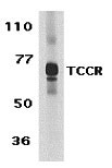
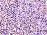
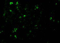
| Application Notes | TCCR antibody can be used for detection of TCCR by Western blot at 1 µg/mL. An approximately 70 kDa band can be detected. Antibody can also be used for immunohistochemistry starting at 5 µg/mL. For immunofluorescence start at 20 µg/mL. |
| Gene ID | 9466 |
|---|---|
| Other Names | TCCR Antibody: CRL1, TCCR, WSX1, IL27R, IL-27RA, zcytor1, CRL1, UNQ296/PRO336, Interleukin-27 receptor subunit alpha, Cytokine receptor WSX-1, IL-27 receptor subunit alpha, interleukin 27 receptor, alpha |
| Target/Specificity | IL27RA; |
| Reconstitution & Storage | TCCR antibody can be stored at 4℃ for three months and -20℃, stable for up to one year. As with all antibodies care should be taken to avoid repeated freeze thaw cycles. Antibodies should not be exposed to prolonged high temperatures. |
| Precautions | TCCR Antibody is for research use only and not for use in diagnostic or therapeutic procedures. |
| Name | IL27RA |
|---|---|
| Synonyms | CRL1, TCCR, WSX1 |
| Function | Receptor for IL27. Requires IL6ST/GP130 to mediate signal transduction in response to IL27. This signaling system acts through STAT3 and STAT1. Acts as a receptor for the neuroprotective peptide humanin as part of a complex with IL6ST/GP130 and CNTFR (PubMed:19386761). Involved in the regulation of Th1-type immune responses. Also appears to be involved in innate defense mechanisms. |
| Cellular Location | Membrane; Single-pass type I membrane protein. |
| Tissue Location | Highly expressed in lymphoid tissues such as spleen, lymph nodes and peripheral blood leukocytes. Weakly expressed in other tissues examined including heart, brain, fetal and adult lung, liver, skeletal muscle, kidney, pancreas, prostate, testis, ovary, small intestine, kidney and colon. In the lymphoid system, higher level expression in CD4+ T-cell subsets than in CD8+ T-cell subsets. Also weaker expression in CD19+ B-cells and monocytes |
For Research Use Only. Not For Use In Diagnostic Procedures.
Provided below are standard protocols that you may find useful for product applications.
BACKGROUND
TCCR Antibody: Upon antigen challenge, T-helper cells differentiate into two functional distinct subsets, Th1 and Th2. Th1 cells produce IL-2, IFN-gamma and lymphotoxin-beta that augment cell mediated immune response while Th2 cells secrete IL-4, IL-5, and IL-10 that enhance humoral immunity. The function of T-helper cells is regulated by cytokines. A novel cytokine receptor was recently identified and cloned. It is a new member in the type I cytokine receptor family and designated TCCR for T-cell cytokine receptor and WSX-1. TCCR deficient mice had impaired Th1 responses to protein antigen challenge, including decreased levels of IFN-gamma and Th1-dependent antibody IgG2a. TCCR is predominately expressed in thymus, spleen, lymph notes and peripheral blood leukocytes.
REFERENCES
Chen Q, Ghilardi N, Wang H, Baker T, Xie MH, Gurney A, Grewal IS and de Sauvage FJ. Development of Th1-type immune responses requires the type I cytokine receptor TCCR Nature 2000;407(6806):916-920
Sprecher,C.A., Grant,F.J., Baumgartner,J.W., Presnell,S.R., Schrader,S.K., Yamagiwa,T., Whitmore,T.E., O'Hara,P.J. and Foster,D.F. Cloning and characterization of a novel class I cytokine receptor Biochem. Biophys. Res. Commun. 1998;246(1):82-90
终于等到您。ABCEPTA(百远生物)抗体产品。
点击下方“我要评价 ”按钮提交您的反馈信息,您的反馈和评价是我们最宝贵的财富之一,
我们将在1-3个工作日内处理您的反馈信息。
如有疑问,联系:0512-88856768 tech-china@abcepta.com.


