


 癌症的基本特征包括细胞增殖、血管生成、迁移、凋亡逃避机制和细胞永生等。找到癌症发生过程中这些通路的关键标记物和对应的抗体用于检测至关重要。
 为您推荐一个泛素化位点预测神器——泛素化分析工具,可以为您的蛋白的泛素化位点作出预测和评分。
 细胞自噬受体图形绘图工具为你的蛋白的细胞受体结合位点作出预测和评分,识别结合到自噬通路中的蛋白是非常重要的,便于让我们理解自噬在正常生理、病理过程中的作用,如发育、细胞分化、神经退化性疾病、压力条件下、感染和癌症。
MAP1 Antibody
- 产品详情
- 实验流程
- 背景知识
Application
| WB, E |
|---|---|
| Primary Accession | Q96BY2 |
| Other Accession | NP_071434, 19923584 |
| Reactivity | Human, Mouse |
| Host | Rabbit |
| Clonality | Polyclonal |
| Isotype | IgG |
| Calculated MW | 39513 Da |
| Concentration (mg/ml) | 1 mg/mL |
| Conjugate | Unconjugated |
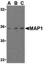
| Application Notes | MAP1 antibody can be used for the detection of MAP-1 by Western blot at 1 to 4 µg/mL. |
| Gene ID | 64112 |
|---|---|
| Other Names | MAP1 Antibody: MAP-1, PNMA4, Modulator of apoptosis 1, Paraneoplastic antigen Ma4, MAP-1, modulator of apoptosis 1 |
| Target/Specificity | MOAP1; |
| Reconstitution & Storage | MAP1 antibody can be stored at 4℃ for three months and -20℃, stable for up to one year. As with all antibodies care should be taken to avoid repeated freeze thaw cycles. Antibodies should not be exposed to prolonged high temperatures. |
| Precautions | MAP1 Antibody is for research use only and not for use in diagnostic or therapeutic procedures. |
| Name | MOAP1 {ECO:0000303|PubMed:19366867, ECO:0000312|HGNC:HGNC:16658} |
|---|---|
| Function | Retrotransposon-derived protein that forms virion-like capsids (By similarity). Acts as an effector of BAX during apoptosis: enriched at outer mitochondria membrane and associates with BAX upon induction of apoptosis, facilitating BAX-dependent mitochondrial outer membrane permeabilization and apoptosis (PubMed:11060313, PubMed:16199525). Required for death receptor-dependent apoptosis (PubMed:11060313). When associated with RASSF1, promotes BAX conformational change and translocation to mitochondrial membranes in response to TNF and TNFSF10 stimulation (PubMed:15949439). Also promotes autophagy: promotes phagophore closure via association with ATG8 proteins (PubMed:33783314). Acts as an inhibitor of the NFE2L2/NRF2 pathway via interaction with SQSTM1: interaction promotes dissociation of SQSTM1 inclusion bodies that sequester KEAP1, relieving inactivation of the BCR(KEAP1) complex (PubMed:33393215). |
| Cellular Location | Cytoplasm, cytosol. Mitochondrion outer membrane Extracellular vesicle membrane {ECO:0000250|UniProtKB:Q9ERH6} Note=Forms virion-like extracellular vesicles that are released from cells. {ECO:0000250|UniProtKB:Q9ERH6} |
| Tissue Location | Widely expressed, with high levels in heart and brain. |
For Research Use Only. Not For Use In Diagnostic Procedures.
Provided below are standard protocols that you may find useful for product applications.
BACKGROUND
MAP1 Antibody: Apoptosis plays a major role in normal organism development, tissue homeostasis, and removal of damaged cells. Disruption of this process has been implicated in a variety of diseases such as cancer. Members of the Bcl-2 family are known to be critical regulators of this process. These proteins are characterized by the presence of several conserved motifs termed Bcl-2 homology (BH) domains. A related protein termed MAP-1 has recently been identified. This protein contains a BH3-like domain and induces caspase-dependent apoptosis in mammalian cells when overexpressed. It forms homodimers and associates with Bcl-2 family members such as Bax, Bcl-2, and Bcl-XL in vitro and in vivo. It has been suggested that MAP-1 associates with the tumor suppressor RASSF1A following death receptor activation, allowing a conformational change in Bax that leads to cellular apoptosis.
REFERENCES
Lockshin RA, Osborne B, and Zakeri Z. Cell death in the third millennium. Cell Death Differ. 2000; 7:2-7.
Cory S, Huang DCS, and Adams JM. The Bcl-2 family: roles in cell survival and oncogenesis. Oncogene 2003; 22:8590-607.
Heiser D, Labi V, Erlacher M, et al. The Bcl-2 protein family and its role in the development of neoplastic disease. Exp. Geron. 2004; 39:1125-35.
Tan KO, Tan KML, Chan S-L, et al. MAP-1, a novel proapoptotic protein containing a BH3-like motif that associates with Bax through its Bcl-2 homology domains. J. Biol. Chem. 2001; 276:2802-7.
终于等到您。ABCEPTA(百远生物)抗体产品。
点击下方“我要评价 ”按钮提交您的反馈信息,您的反馈和评价是我们最宝贵的财富之一,
我们将在1-3个工作日内处理您的反馈信息。
如有疑问,联系:0512-88856768 tech-china@abcepta.com.


