


 癌症的基本特征包括细胞增殖、血管生成、迁移、凋亡逃避机制和细胞永生等。找到癌症发生过程中这些通路的关键标记物和对应的抗体用于检测至关重要。
 为您推荐一个泛素化位点预测神器——泛素化分析工具,可以为您的蛋白的泛素化位点作出预测和评分。
 细胞自噬受体图形绘图工具为你的蛋白的细胞受体结合位点作出预测和评分,识别结合到自噬通路中的蛋白是非常重要的,便于让我们理解自噬在正常生理、病理过程中的作用,如发育、细胞分化、神经退化性疾病、压力条件下、感染和癌症。
PAK5 Antibody
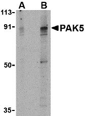
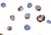
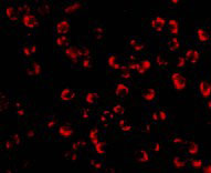
- 产品详情
- 实验流程
- 背景知识
Application 
| WB, IF, ICC, E |
|---|---|
| Primary Accession | Q9P286 |
| Other Accession | Q9P286, 12585290 |
| Reactivity | Human |
| Host | Rabbit |
| Clonality | Polyclonal |
| Isotype | IgG |
| Calculated MW | 80745 Da |
| Concentration (mg/ml) | 1 mg/mL |
| Conjugate | Unconjugated |
| Application Notes | PAK5 antibody can be used for the detection of PAK5 by Western blot at 2 - 4 µg/mL. Antibody can also be used for immunocytochemistry starting at 2 µg/mL. For immunofluorescence start at 10 µg/mL. |
| Gene ID | 57144 |
|---|---|
| Other Names | PAK5 Antibody: PAK5, KIAA1264, PAK5, p21-activated kinase 5, PAK-5, p21 protein (Cdc42/Rac)-activated kinase 7 |
| Target/Specificity | PAK7; |
| Reconstitution & Storage | PAK5 antibody can be stored at 4℃ for three months and -20℃, stable for up to one year. As with all antibodies care should be taken to avoid repeated freeze thaw cycles. Antibodies should not be exposed to prolonged high temperatures. |
| Precautions | PAK5 Antibody is for research use only and not for use in diagnostic or therapeutic procedures. |
For Research Use Only. Not For Use In Diagnostic Procedures.
| Name | PAK5 (HGNC:15916) |
|---|---|
| Synonyms | KIAA1264, PAK7 |
| Function | Serine/threonine protein kinase that plays a role in a variety of different signaling pathways including cytoskeleton regulation, cell migration, proliferation or cell survival. Activation by various effectors including growth factor receptors or active CDC42 and RAC1 results in a conformational change and a subsequent autophosphorylation on several serine and/or threonine residues. Phosphorylates the proto-oncogene RAF1 and stimulates its kinase activity. Promotes cell survival by phosphorylating the BCL2 antagonist of cell death BAD. Phosphorylates CTNND1, probably to regulate cytoskeletal organization and cell morphology. Keeps microtubules stable through MARK2 inhibition and destabilizes the F-actin network leading to the disappearance of stress fibers and focal adhesions. |
| Cellular Location | Mitochondrion. Cytoplasm. Nucleus. Note=Shuttles between the nucleus and the mitochondria, and mitochondrial localization is essential for the role in cell survival |
| Tissue Location | Predominantly expressed in brain. |
Provided below are standard protocols that you may find useful for product applications.
BACKGROUND
PAK5 Antibody: The p21-activated kinases (PAKs) are serine-threonine kinases that bind to the active forms of Cdc42 and Rac. They are divided into two groups, the first of which include PAK1, 2 and 3, and can be activated by Cdc42/Rac binding. Group 1 PAKs contain an autoinhibitory domain whose activity is regulated by Cdc42/Rac binding. The group 1 PAKs are known to be involved in cellular processes such as gene transcription, apoptosis, and cell morphology and motility. Much less is known about the second group, which includes PAK4, 5 and 6. These proteins are not activated by Cdc42/Rac binding. PAK5 was initially identified as a kinase expressed primarily in brain that while possessing a kinase domain and GTPase binding domain similar to PAK4 and PAK6, is completely different from both. Expression of PAK5 in neural based cell lines resulted in neurite outgrowth suggesting that PAK5 may be involved in regulating the cytoskeletal changes necessary for promoting neurite outgrowth. Other experiments suggest that unlike the other PAKs, PAK5 may inhibit apoptosis by phosphorylating the Bcl-2 family member Bad.
REFERENCES
Jaffer ZM and Chernoff J. p21-activated kinases: three more join the Pak. Int. J. Biochem. Cell Biol. 2002; 34:713-7.
Dan C, Nath N, Liberto M, et al. PAK5, a new brain-specific kinase, promotes neurite outgrowth in N1E-115 cells. Mol. Cell. Biol. 2002; 22:567-77.
Cotteret S, Jaffer ZM, Beeser A, et al. p21-activated kinase 5 (Pak5) localizes to mitochondria and inhibits apoptosis by phosphorylated BAD. Mol. Cell. Biol. 2003; 23:5526-39.

终于等到您。ABCEPTA(百远生物)抗体产品。
点击下方“我要评价 ”按钮提交您的反馈信息,您的反馈和评价是我们最宝贵的财富之一,
我们将在1-3个工作日内处理您的反馈信息。
如有疑问,联系:0512-88856768 tech-china@abcepta.com.


