


 癌症的基本特征包括细胞增殖、血管生成、迁移、凋亡逃避机制和细胞永生等。找到癌症发生过程中这些通路的关键标记物和对应的抗体用于检测至关重要。
 为您推荐一个泛素化位点预测神器——泛素化分析工具,可以为您的蛋白的泛素化位点作出预测和评分。
 细胞自噬受体图形绘图工具为你的蛋白的细胞受体结合位点作出预测和评分,识别结合到自噬通路中的蛋白是非常重要的,便于让我们理解自噬在正常生理、病理过程中的作用,如发育、细胞分化、神经退化性疾病、压力条件下、感染和癌症。
ATR Antibody
- 产品详情
- 实验流程
- 背景知识
Application
| WB, IF, E, IHC-P |
|---|---|
| Primary Accession | Q9H6X2 |
| Other Accession | NP_444262, 16933551 |
| Reactivity | Human, Mouse, Rat |
| Host | Rabbit |
| Clonality | Polyclonal |
| Isotype | IgG |
| Calculated MW | 62789 Da |
| Concentration (mg/ml) | 1 mg/mL |
| Conjugate | Unconjugated |
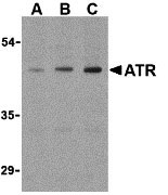
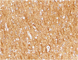
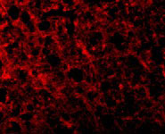
| Application Notes | ATR antibody can be used for detection of ATR by Western blot at 0.5 to 2 µg/mL. Antibody can also be used for immunohistochemistry starting at 2 µg/mL. For immunofluorescence start at 10 µg/mL. |
| Gene ID | 84168 |
|---|---|
| Other Names | ATR Antibody: ATR, GAPO, TEM8, ATR, Anthrax toxin receptor 1, Tumor endothelial marker 8, anthrax toxin receptor 1 |
| Target/Specificity | ANTXR1; At least three isoforms of ATR are known to exist; this antibody will detect all three isoforms. |
| Reconstitution & Storage | ATR antibody can be stored at 4℃ for three months and -20℃, stable for up to one year. As with all antibodies care should be taken to avoid repeated freeze thaw cycles. Antibodies should not be exposed to prolonged high temperatures. |
| Precautions | ATR Antibody is for research use only and not for use in diagnostic or therapeutic procedures. |
| Name | ANTXR1 {ECO:0000303|PubMed:22912819, ECO:0000312|HGNC:HGNC:21014} |
|---|---|
| Function | Plays a role in cell attachment and migration. Interacts with extracellular matrix proteins and with the actin cytoskeleton and thereby plays an important role in normal extracellular matrix (ECM) homeostasis. Mediates adhesion of cells to type 1 collagen and gelatin, reorganization of the actin cytoskeleton and promotes cell spreading. Plays a role in the angiogenic response of cultured umbilical vein endothelial cells. May also act as a receptor for PLAU. Upon ligand binding, stimulates the phosphorylation of EGFR and ERK1/2 (PubMed:30241478). |
| Cellular Location | Cell membrane; Single-pass type I membrane protein. Cell projection, lamellipodium membrane; Single-pass type I membrane protein. Cell projection, filopodium membrane; Single-pass type I membrane protein. Note=At the membrane of lamellipodia and at the tip of actin-enriched filopodia (PubMed:16762926). Colocalizes with actin at the base of lamellipodia (PubMed:16762926) |
| Tissue Location | Detected in umbilical vein endothelial cells (at protein level). Highly expressed in tumor endothelial cells |
For Research Use Only. Not For Use In Diagnostic Procedures.
Provided below are standard protocols that you may find useful for product applications.
BACKGROUND
ATR Antibody: The Anthrax toxin receptor (ATR) was initially discovered as the tumor endothelial marker 8 (TEM8). This protein, which exists in three isoforms (36, 40, and 60 kDa), is highly expressed in tumor vessels as well as in the vasculature of developing embryos, suggesting that it may normally play a role in angiogenesis. However, it also acts as the receptor for anthrax toxin. Following the binding of this protein by the protective antigen (PA) of anthrax, PA is cleaved and heptamerizes to form the binding site for both edema factor (EF) and lethal factor (LF). This complex is then endocytosed by the cell; acidification in endosomes allows the release of EF and LF into the cytoplasm where they interfere with MAPK signaling and induce apoptosis.
REFERENCES
Carson-Walter EB, Watkins DN, Nanda A, et al. Cell surface tumor endothelial markers are conserved in mice and humans. Can. Res. 2001; 61:6649-6655.
Bradley KA, Mogridge J, Mourez M, et al. Identification of the cellular receptor for anthrax toxin. Nature 2001; 414:225-9.
Molloy S, Bresnahan PA, Thomas G, et al. Human furin is a calcium-dependent serine endoprotease that recognizes the sequence Arg-X-X-Arg and efficiently cleaves anthrax toxin protective antigen. J. Biol. Chem. 1992; 267:16396-402.
Duesbery N, Webb C, Vande Woude G, et al. Proteolytic inactivation of MAP-kinase-kinase by anthrax lethal factor. Science 1998; 280:734-6.
终于等到您。ABCEPTA(百远生物)抗体产品。
点击下方“我要评价 ”按钮提交您的反馈信息,您的反馈和评价是我们最宝贵的财富之一,
我们将在1-3个工作日内处理您的反馈信息。
如有疑问,联系:0512-88856768 tech-china@abcepta.com.


