


 癌症的基本特征包括细胞增殖、血管生成、迁移、凋亡逃避机制和细胞永生等。找到癌症发生过程中这些通路的关键标记物和对应的抗体用于检测至关重要。
 为您推荐一个泛素化位点预测神器——泛素化分析工具,可以为您的蛋白的泛素化位点作出预测和评分。
 细胞自噬受体图形绘图工具为你的蛋白的细胞受体结合位点作出预测和评分,识别结合到自噬通路中的蛋白是非常重要的,便于让我们理解自噬在正常生理、病理过程中的作用,如发育、细胞分化、神经退化性疾病、压力条件下、感染和癌症。
IRAK-4 Antibody
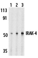
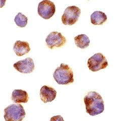
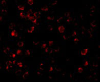
- 产品详情
- 实验流程
- 背景知识
Application 
| WB, IF, ICC, E |
|---|---|
| Primary Accession | Q9NWZ3 |
| Other Accession | AAM15772, 20219010 |
| Reactivity | Human |
| Host | Rabbit |
| Clonality | Polyclonal |
| Isotype | IgG |
| Calculated MW | 51530 Da |
| Concentration (mg/ml) | 1 mg/mL |
| Conjugate | Unconjugated |
| Application Notes | IRAK-4 antibody can be used for the detection of IRAK-4 by Western blot at 1 to 4 µg/mL. Antibody can also be used for immunocytochemistry starting at 10 µg/mL. For immunofluorescence start at 10 µg/mL. |
| Gene ID | 51135 |
|---|---|
| Other Names | IRAK-4 Antibody: IPD1, REN64, IRAK-4, NY-REN-64, Interleukin-1 receptor-associated kinase 4, Renal carcinoma antigen NY-REN-64, interleukin-1 receptor-associated kinase 4 |
| Target/Specificity | IRAK4; IRAK-4 antibody is predicted to not cross-react with other members of the IRAK protein family. |
| Reconstitution & Storage | IRAK-4 antibody can be stored at 4℃ for three months and -20℃, stable for up to one year. As with all antibodies care should be taken to avoid repeated freeze thaw cycles. Antibodies should not be exposed to prolonged high temperatures. |
| Precautions | IRAK-4 Antibody is for research use only and not for use in diagnostic or therapeutic procedures. |
For Research Use Only. Not For Use In Diagnostic Procedures.
| Name | IRAK4 |
|---|---|
| Function | Serine/threonine-protein kinase that plays a critical role in initiating innate immune response against foreign pathogens. Involved in Toll-like receptor (TLR) and IL-1R signaling pathways (PubMed:17878374). Is rapidly recruited by MYD88 to the receptor- signaling complex upon TLR activation to form the Myddosome together with IRAK2. Phosphorylates initially IRAK1, thus stimulating the kinase activity and intensive autophosphorylation of IRAK1. Phosphorylates E3 ubiquitin ligases Pellino proteins (PELI1, PELI2 and PELI3) to promote pellino-mediated polyubiquitination of IRAK1. Then, the ubiquitin- binding domain of IKBKG/NEMO binds to polyubiquitinated IRAK1 bringing together the IRAK1-MAP3K7/TAK1-TRAF6 complex and the NEMO-IKKA-IKKB complex. In turn, MAP3K7/TAK1 activates IKKs (CHUK/IKKA and IKBKB/IKKB) leading to NF-kappa-B nuclear translocation and activation. Alternatively, phosphorylates TIRAP to promote its ubiquitination and subsequent degradation. Phosphorylates NCF1 and regulates NADPH oxidase activation after LPS stimulation suggesting a similar mechanism during microbial infections. |
| Cellular Location | Cytoplasm. |
Provided below are standard protocols that you may find useful for product applications.
BACKGROUND
IRAK-4 Antibody: Interleukin-1 (IL-1) and lipopolysaccharide (LPS) induces cellular responses through IL-1 receptor (IL-1R) and Toll-like receptors (TLR). IL-1R-associated kinases (IRAK, IRAK2, and IRAK-M) regulate the activation of NF-κB and MAP kinase (MAPK) by IL-1R/TLR. A novel member in the IRAK/Pelle family was recently identified and designated IRAK-4. Overexpression of IRAK-4 activates NF-κB and MAPK pathways. IRAK-4 interacts with and phosphorylates IRAK-1. IRAK-4-deficient animals are completely resistant to the challenge with LPS. Animals and humans lacking IRAK-4 are impaired in their responses to viral and bacterial challenges. Members in IRAK/Pelle family play a central role in IL-1R/TLR mediated inflammatory responses and in innate immunity.
REFERENCES
Cao Z, Henzel WJ, and Gao X. IRAK: a kinase associated with the interleukin-1 receptor. Science 1996; 271:1128-31.
Muzio M, Ni J, Feng P, et al. IRAK (Pelle) family member IRAK-2 and MyD88 as proximal mediators of IL-1 signaling. Science 1997; 278:1612-5
Wesche H, Gao X, Li X, et al. IRAK-M is a novel member of the Pelle/interleukin-1 receptor-associated kinase (IRAK) family. J. Biol. Chem. 1999; 274:19403-10.
Li S, Strelow A, Fontana EJ, et al. IRAK-4: a novel member of the IRAK family with the properties of an IRAK-kinase. Proc. Natl. Acad. Sci. USA 2002; 99:5567-72.

终于等到您。ABCEPTA(百远生物)抗体产品。
点击下方“我要评价 ”按钮提交您的反馈信息,您的反馈和评价是我们最宝贵的财富之一,
我们将在1-3个工作日内处理您的反馈信息。
如有疑问,联系:0512-88856768 tech-china@abcepta.com.


