


 癌症的基本特征包括细胞增殖、血管生成、迁移、凋亡逃避机制和细胞永生等。找到癌症发生过程中这些通路的关键标记物和对应的抗体用于检测至关重要。
 为您推荐一个泛素化位点预测神器——泛素化分析工具,可以为您的蛋白的泛素化位点作出预测和评分。
 细胞自噬受体图形绘图工具为你的蛋白的细胞受体结合位点作出预测和评分,识别结合到自噬通路中的蛋白是非常重要的,便于让我们理解自噬在正常生理、病理过程中的作用,如发育、细胞分化、神经退化性疾病、压力条件下、感染和癌症。
Bcl-B Antibody
- 产品详情
- 实验流程
- 背景知识
Application
| WB, ICC, E |
|---|---|
| Primary Accession | Q9HD36 |
| Other Accession | NP_065129, 9966783 |
| Reactivity | Human |
| Host | Rabbit |
| Clonality | Polyclonal |
| Isotype | IgG |
| Calculated MW | 23204 Da |
| Concentration (mg/ml) | 1 mg/mL |
| Conjugate | Unconjugated |
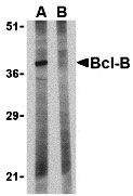
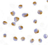
| Application Notes | Bcl-B antibody can be used for the detection of Bcl-B by Western blot at 1 µg/mL. Despite its predicted molecular weight, Bcl-B is often at higher molecular weights, presumably due to post-translational modifications. Antibody can also be used for immunocytochemistry starting at 10 µg/mL. |
| Gene ID | 10017 |
|---|---|
| Other Names | Bcl-B Antibody: Boo, Diva, BCL-B, BCLB, Bcl-2-like protein 10, Anti-apoptotic protein NrH, Bcl2-L-10, BCL2-like 10 (apoptosis facilitator) |
| Target/Specificity | BCL2L10; |
| Reconstitution & Storage | Bcl-B antibody can be stored at 4℃ for three months and -20℃, stable for up to one year. As with all antibodies care should be taken to avoid repeated freeze thaw cycles. Antibodies should not be exposed to prolonged high temperatures. |
| Precautions | Bcl-B Antibody is for research use only and not for use in diagnostic or therapeutic procedures. |
| Name | BCL2L10 {ECO:0000303|PubMed:17532299} |
|---|---|
| Function | Promotes cell survival by suppressing apoptosis induced by BAX but not BAK (PubMed:11278245, PubMed:11689480). Increases binding of AHCYL1/IRBIT to ITPR1 (PubMed:27995898). Reduces ITPR1-mediated calcium release from the endoplasmic reticulum cooperatively with AHCYL1/IRBIT under normal cellular conditions (PubMed:27995898). Under apoptotic stress conditions, dissociates from ITPR1 and is displaced from mitochondria-associated endoplasmic reticulum membranes, leading to increased Ca(2+) transfer to mitochondria which promotes apoptosis (PubMed:27995898). Required for the correct formation of the microtubule organizing center during oocyte cell division, potentially via regulation of protein abundance and localization of other microtubule organizing center components such as AURKA and TPX2 (By similarity). |
| Cellular Location | Mitochondrion. Nucleus membrane. Endoplasmic reticulum. Cytoplasm, cytoskeleton, spindle {ECO:0000250|UniProtKB:Q9Z0F3}. Note=Localizes to mitochondria-associated endoplasmic reticulum membranes (MAMs) (PubMed:27995898). Localization to MAMs is greatly reduced under apoptotic stress conditions (PubMed:27995898) |
| Tissue Location | Widely expressed in adult tissues. Preferentially expressed in lung, liver and kidney. |
For Research Use Only. Not For Use In Diagnostic Procedures.
Provided below are standard protocols that you may find useful for product applications.
BACKGROUND
Bcl-B Antibody: Members in the Bcl-2 family are critical regulators of apoptosis by either inhibiting or promoting cell death. Bcl-B is a recently discovered anti-apoptotic member of the Bcl-2. Unlike the mouse homolog (also known as Diva/Boo) which is predominantly expressed in ovary and testis, the human Bcl-B protein is widely expressed. Also, the human Bcl-B functions by binding to and suppressing the apoptotic activity of Bax, whereas the mouse homolog binds Bak and also interacts with the apoptosis protein Apaf-1.
REFERENCES
Cory S, Huang DCS, and Adams JM. The Bcl-2 family: roles in cell survival and oncogenesis. Oncogene 2003; 22:8590-607
Heiser D, Labi V, Erlacher M, et al. The Bcl-2 protein family and its role in the development of neoplastic disease. Exp. Geron. 2004; 39:1125-35.
Ke N, Godzik A, and Reed JC. Bcl-B: A novel Bcl-2 family member that differentially binds and regulates Bax and Bak. J. Biol. Chem. 2001; 276:12481-4.
Song Q, Kuang Y, Dixit VM, et al. Boo, a negative regulator of cell death, interacts with Apaf-1. EMBO J. 1999; 18:167-78.
终于等到您。ABCEPTA(百远生物)抗体产品。
点击下方“我要评价 ”按钮提交您的反馈信息,您的反馈和评价是我们最宝贵的财富之一,
我们将在1-3个工作日内处理您的反馈信息。
如有疑问,联系:0512-88856768 tech-china@abcepta.com.


