


 癌症的基本特征包括细胞增殖、血管生成、迁移、凋亡逃避机制和细胞永生等。找到癌症发生过程中这些通路的关键标记物和对应的抗体用于检测至关重要。
 为您推荐一个泛素化位点预测神器——泛素化分析工具,可以为您的蛋白的泛素化位点作出预测和评分。
 细胞自噬受体图形绘图工具为你的蛋白的细胞受体结合位点作出预测和评分,识别结合到自噬通路中的蛋白是非常重要的,便于让我们理解自噬在正常生理、病理过程中的作用,如发育、细胞分化、神经退化性疾病、压力条件下、感染和癌症。
XAF-1 Antibody
- 产品详情
- 实验流程
- 背景知识
Application
| WB, IF, E |
|---|---|
| Primary Accession | Q6GPH4 |
| Other Accession | CAA68030, 1869901 |
| Reactivity | Human |
| Host | Rabbit |
| Clonality | Polyclonal |
| Isotype | IgG |
| Calculated MW | 34626 Da |
| Concentration (mg/ml) | 1 mg/mL |
| Conjugate | Unconjugated |
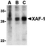
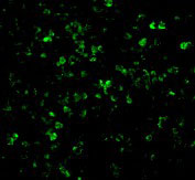
| Application Notes | XAF-1 antibody can be used for the detection of XAF-1 by Western blot at 1 - 4 µg/mL. Multiple isoforms of XAF-1 exist and a doublet at ~30 kDa can be seen using anti-XAF-1 (IN). For immunofluorescence start at 20 µg/mL. |
| Gene ID | 54739 |
|---|---|
| Other Names | XAF-1 Antibody: BIRC4BP, XIAPAF1, HSXIAPAF1, BIRC4BP, XIAP-associated factor 1, BIRC4-binding protein, XIAP associated factor 1 |
| Target/Specificity | XAF1; |
| Reconstitution & Storage | XAF-1antibody can be stored at 4℃ for three months and -20℃, stable for up to one year. As with all antibodies care should be taken to avoid repeated freeze thaw cycles. Antibodies should not be exposed to prolonged high temperatures. |
| Precautions | XAF-1 Antibody is for research use only and not for use in diagnostic or therapeutic procedures. |
| Name | XAF1 |
|---|---|
| Synonyms | BIRC4BP, XIAPAF1 |
| Function | Seems to function as a negative regulator of members of the IAP (inhibitor of apoptosis protein) family. Inhibits anti-caspase activity of BIRC4. Induces cleavage and inactivation of BIRC4 independent of caspase activation. Mediates TNF-alpha-induced apoptosis and is involved in apoptosis in trophoblast cells. May inhibit BIRC4 indirectly by activating the mitochondrial apoptosis pathway. After translocation to mitochondria, promotes translocation of BAX to mitochondria and cytochrome c release from mitochondria. Seems to promote the redistribution of BIRC4 from the cytoplasm to the nucleus, probably independent of BIRC4 inactivation which seems to occur in the cytoplasm. The BIRC4-XAF1 complex mediates down-regulation of BIRC5/survivin; the process requires the E3 ligase activity of BIRC4. Seems to be involved in cellular sensitivity to the proapoptotic actions of TRAIL. May be a tumor suppressor by mediating apoptosis resistance of cancer cells. |
| Cellular Location | Cytoplasm. Nucleus. Mitochondrion. Note=Found in the cytoplasm and nucleus of placental syncytiotrophoblasts Translocates to mitochondria upon TNF-alpha treatment [Isoform 5]: Nucleus. |
| Tissue Location | Widely expressed. Expression is frequently down- regulated in cancer cell lines. Isoform 5 is widely expressed Expressed in placenta (at protein level). |
For Research Use Only. Not For Use In Diagnostic Procedures.
Provided below are standard protocols that you may find useful for product applications.
BACKGROUND
XAF-1 Antibody: XAF-1 binds to XIAP, an inhibitor of caspases-3, -7, and -9, and triggers its relocation from the cytosol to the nucleus. Overexpression of XAF-1 results in the neutralization of XIAP's ability to inhibit cell death. XAF-1 is normally expressed in all adult and fetal tissues but was found to be present in very low levels in a variety of cancer cell lines. In contrast, XIAP levels have been shown to be high in a majority of cell lines. Low XAF-1 and high basal expression of XIAP may therefore play a critical role in maintaining survival of cancer cell lines. Both IFN-alpha2 and IFN-beta can induce XAF-1 mRNA in all cells examined but induction of XAF-1 protein (as observed by immunoblot analysis) was seen only in cell lines sensitive to the apoptotic effects of IFNs.
REFERENCES
Liston P, Fong W, Kelly NL, et al. Identification of XAF1 as an antagonist of XIAP anticaspase activity. Nature Cell Biol. 2001;3:128-33.
Deveraux QL, Takahashi R, Savesan GS, and Reed JC. X-linked IAP is a direct inhibitor of cell-death proteases. Nature 1997;388:300-4.
Fong WG, Liston P, Rajcan-Separovic E, et al. Expression and genetic analysis of XIAP-associated factor 1 (XAF1) in cancer cell lines. Genomics 2000;70:113-122.
Leaman DW, Chawla-Sarkar M, Vyas K, et al. Identification of X-linked inhibitor of apoptosis associated factor-1 as an interferon-stimulated gene that augments TRAIL/Apo2L-induced apoptosis. J. Biol. Chem. 2002;277:28504-11.
终于等到您。ABCEPTA(百远生物)抗体产品。
点击下方“我要评价 ”按钮提交您的反馈信息,您的反馈和评价是我们最宝贵的财富之一,
我们将在1-3个工作日内处理您的反馈信息。
如有疑问,联系:0512-88856768 tech-china@abcepta.com.


