


 癌症的基本特征包括细胞增殖、血管生成、迁移、凋亡逃避机制和细胞永生等。找到癌症发生过程中这些通路的关键标记物和对应的抗体用于检测至关重要。
 为您推荐一个泛素化位点预测神器——泛素化分析工具,可以为您的蛋白的泛素化位点作出预测和评分。
 细胞自噬受体图形绘图工具为你的蛋白的细胞受体结合位点作出预测和评分,识别结合到自噬通路中的蛋白是非常重要的,便于让我们理解自噬在正常生理、病理过程中的作用,如发育、细胞分化、神经退化性疾病、压力条件下、感染和癌症。
T-cadherin Antibody
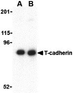
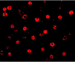
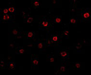
- 产品详情
- 实验流程
- 背景知识
Application 
| WB, IF, E |
|---|---|
| Primary Accession | O75355 |
| Other Accession | NP_001248, 166197701 |
| Reactivity | Human, Mouse |
| Host | Rabbit |
| Clonality | Polyclonal |
| Isotype | IgG |
| Calculated MW | 59105 Da |
| Concentration (mg/ml) | 1 mg/mL |
| Conjugate | Unconjugated |
| Application Notes | T-cadherin antibody can be used for the detection of T-cadherin by Western blot at 0.5 - 1 µg/mL. Antibody can also be used for immunocytochemistry starting at 20 µg/mL. For immunofluorescence start at 20 µg/mL. |
| Gene ID | 956 |
|---|---|
| Other Names | T-cadherin Antibody: HB6, CD39L3, NTPDase-3, Ectonucleoside triphosphate diphosphohydrolase 3, CD39 antigen-like 3, NTPDase 3, ectonucleoside triphosphate diphosphohydrolase 3 |
| Target/Specificity | ENTPD3; At least two isoforms of T-cadherin are known to exist. |
| Reconstitution & Storage | T-cadherin antibody can be stored at 4℃ for three months and -20℃, stable for up to one year. As with all antibodies care should be taken to avoid repeated freeze thaw cycles. Antibodies should not be exposed to prolonged high temperatures. |
| Precautions | T-cadherin Antibody is for research use only and not for use in diagnostic or therapeutic procedures. |
For Research Use Only. Not For Use In Diagnostic Procedures.
| Name | ENTPD3 (HGNC:3365) |
|---|---|
| Function | Catalyzes the hydrolysis of nucleoside triphosphates (NTPs) and diphosphates (NDPs) (PubMed:10231536, PubMed:11300774, PubMed:15966724, PubMed:9675246). The enzyme sequentially removes phosphate groups in two successive steps, converting NTPs to nucleoside monophosphates (NMPs) via NDP intermediates (PubMed:10231536, PubMed:11300774, PubMed:15966724, PubMed:9675246). This activity contributes to the regulation of extracellular levels of nucleotides (PubMed:10231536, PubMed:11300774, PubMed:15966724, PubMed:9675246). Has a preference for the hydrolysis of ATP (PubMed:10231536). Does not hydrolyzes AMP (PubMed:9675246). |
| Cellular Location | Cell membrane; Multi-pass membrane protein |
| Tissue Location | Expressed in adult brain, pancreas, spleen and prostate (PubMed:9676430). Moderate or low expression is seen in most tissues (PubMed:9676430). Not expressed in liver and peripheral blood leukocytes (PubMed:9676430). |
Provided below are standard protocols that you may find useful for product applications.
BACKGROUND
T-cadherin Antibody: T-cadherin was initially identified as cadherin-type cell adhesion molecule expressed in various neuronal populations in a temporally and spatially restricted pattern during axon growth. T-cadherin is an atypical member of the cadherin family because it does not possess the typical transmembrane and cytoplasmic domains but is instead anchored to the plasma membrane by glycosylphosphatidylinositol (GPI) linkage. T-cadherin may play a role in malignant tumor development as loss of the chromosome locus containing the T-cadherin gene correlates with the development of a variety of cancers. Recently it has been shown that T-cadherin can act as a receptor for hexameric and high-molecular weight forms of adiponectin, suggesting that T-cadherin may also play a role in metabolic regulation.
REFERENCES
Ranscht B and Dours-Zimmerman MT. T-cadherin, a novel cadherin cell adhesion molecule in the nervous system lacks the conserved cytoplasmic region. Neuron 1991; 7:391-402.
Ivanov DB, Philippova MP, and Tkachuk VA. Structure and functions of classical cadherins Biochemistry (Moscow) 2001; 66:1175-66.
Takeuchi T, Misaki A, Chen BK, et al. H-cadherin expression in breast cancer. Histopathology 1999; 35:87-88.
Sato M, Mori Y, Sakurada A, et al. The H-cadherin (CDH13) gene is inactivated in human lung cancer. Hum. Gen. 1998; 103:96-101.

终于等到您。ABCEPTA(百远生物)抗体产品。
点击下方“我要评价 ”按钮提交您的反馈信息,您的反馈和评价是我们最宝贵的财富之一,
我们将在1-3个工作日内处理您的反馈信息。
如有疑问,联系:0512-88856768 tech-china@abcepta.com.


