


 癌症的基本特征包括细胞增殖、血管生成、迁移、凋亡逃避机制和细胞永生等。找到癌症发生过程中这些通路的关键标记物和对应的抗体用于检测至关重要。
 为您推荐一个泛素化位点预测神器——泛素化分析工具,可以为您的蛋白的泛素化位点作出预测和评分。
 细胞自噬受体图形绘图工具为你的蛋白的细胞受体结合位点作出预测和评分,识别结合到自噬通路中的蛋白是非常重要的,便于让我们理解自噬在正常生理、病理过程中的作用,如发育、细胞分化、神经退化性疾病、压力条件下、感染和癌症。
IL-31 Antibody

- 产品详情
- 实验流程
- 背景知识
Application 
| WB, IF, E |
|---|---|
| Primary Accession | Q6EAL8 |
| Other Accession | AAS82846, 46241139 |
| Reactivity | Human |
| Host | Rabbit |
| Clonality | Polyclonal |
| Isotype | IgG |
| Calculated MW | 18120 Da |
| Concentration (mg/ml) | 1 mg/mL |
| Conjugate | Unconjugated |
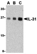
| Application Notes | IL-31 antibody can be used for the detection of IL-31 by Western blot at 2.5 - 10 µg/mL. Despite its predicted size, IL-31 runs at approximately 27 - 30 kDa in SDS-PAGE. For immunofluorescence start at 20 µg/mL. |
| Gene ID | 76399 |
|---|---|
| Other Names | IL-31 Antibody: 1700013B14Rik, Interleukin-31, IL-31, interleukin 31 |
| Target/Specificity | Il31; |
| Reconstitution & Storage | IL-31 antibody can be stored at 4℃ for three months and -20℃, stable for up to one year. As with all antibodies care should be taken to avoid repeated freeze thaw cycles. Antibodies should not be exposed to prolonged high temperatures. |
| Precautions | IL-31 Antibody is for research use only and not for use in diagnostic or therapeutic procedures. |
For Research Use Only. Not For Use In Diagnostic Procedures.
| Name | Il31 {ECO:0000312|MGI:MGI:1923649} |
|---|---|
| Function | Functions as a cytokine that binds to IL31RA, a membrane- bound signal-transducing subunit of the IL31 receptor complex (heterodimer composed of OSMR and IL31RA) (By similarity). Functionally, IL31 signaling via OSMR/IL31RA is particularly important in skin immunity (PubMed:15184896). Mechanistically, ligand binding to IL31RA, induces heterodimerization with OSMR, activating JAK1 and JAK2 (and to a lesser extent TYK2) associated with the intracellular domains of IL31RA and OSMR. These kinases phosphorylate tyrosine residues on IL31RA and OSMR, creating docking sites for STAT1, STAT3, and STAT5B, which are then phosphorylated and activated. In addition, the IL31 receptor complex activates the MAPK pathway (MAPK3/ERK1-MAPK1/ERK2) via recruitment of the adapter protein SHC1 (By similarity). Enhances myeloid progenitor cell survival in vitro (PubMed:17379091). Induces RETNLA and serum amyloid A protein expression in macrophages (PubMed:25847241). |
| Cellular Location | Secreted {ECO:0000250|UniProtKB:Q6EBC2}. |
Provided below are standard protocols that you may find useful for product applications.
BACKGROUND
IL-31 Antibody: Interleukin-31 (IL-31) is a recently discovered T-cell cytokine closely related to IL-6 type cytokines and is preferentially produced by T helper type 2 cells. IL-31 activity is mediated through the ligand-induced oligomerization of a dimeric receptor complex containing IL-31 receptor A and oncostatin M receptor. In response to IL-31 binding, these proteins activate the JAK/STAT and the AKT signaling pathways. RNA levels of IL-31 receptor A and oncostatin M receptor are induced in activated monocytes but are expressed constitutively in epithelial cells. IL-31, when overexpressed in transgenic mice, results in the development of pruritis, alopecia, and skin lesions and in humans may result in atopic dermatitis, suggesting that IL-31 may represent a novel target for antipruritic drug development.
REFERENCES
Dillon SR, Sprecher C, Hammond A, et al. Interleukin 31, a cytokine produced by activated T cells, induces dermatitis in mice. Nat. Immunol. 2004; 5:752-60.
Dreuw A, Radtke S, Pflanz S, et al. Characterization of the signaling capacities of the novel gp130-like cytokine receptor. J. Biol. Chem. 2004; 279:36112-20.
Diveu C, Lak-Hal AH, Froger J, et al. Predominant expression of the long isoform of GP130-like (GPL) receptor is required for interleukin-31 signaling. Eur. Cytokine Netw. 2004; 15:291-302.
Bilsborough J, Leung DY, Maurer M, et al. IL-31 is associated with cutaneous lymphocyte antigen-positive skin homing T cells in patients with atopic dermatitis. J. Allergy Clin. Immunol. 2006; 117:418-25.

终于等到您。ABCEPTA(百远生物)抗体产品。
点击下方“我要评价 ”按钮提交您的反馈信息,您的反馈和评价是我们最宝贵的财富之一,
我们将在1-3个工作日内处理您的反馈信息。
如有疑问,联系:0512-88856768 tech-china@abcepta.com.


