


 癌症的基本特征包括细胞增殖、血管生成、迁移、凋亡逃避机制和细胞永生等。找到癌症发生过程中这些通路的关键标记物和对应的抗体用于检测至关重要。
 为您推荐一个泛素化位点预测神器——泛素化分析工具,可以为您的蛋白的泛素化位点作出预测和评分。
 细胞自噬受体图形绘图工具为你的蛋白的细胞受体结合位点作出预测和评分,识别结合到自噬通路中的蛋白是非常重要的,便于让我们理解自噬在正常生理、病理过程中的作用,如发育、细胞分化、神经退化性疾病、压力条件下、感染和癌症。
TIM-1 Antibody
- 产品详情
- 实验流程
- 背景知识
Application
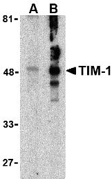
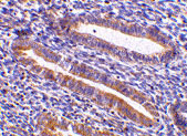
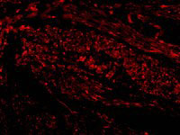
| WB, IF, E, IHC-P |
|---|---|
| Primary Accession | Q96D42 |
| Other Accession | NP_036338, 153085427 |
| Reactivity | Human, Mouse |
| Host | Rabbit |
| Clonality | Polyclonal |
| Isotype | IgG |
| Calculated MW | 39250 Da |
| Concentration (mg/ml) | 1 mg/mL |
| Conjugate | Unconjugated |
| Application Notes | TIM-1 antibody can be used for the detection of TIM-1 by Western blot at 1 - 2 µg/mL. Antibody can also be used for immunohistochemistry starting at 10 µg/mL. For immunofluorescence start at 20 µg/mL. |
| Gene ID | 26762 |
|---|---|
| Other Names | TIM-1 Antibody: TIM, KIM1, TIM1, HAVCR, KIM-1, TIM-1, TIMD1, TIMD-1, HAVCR-1, Hepatitis A virus cellular receptor 1, Kidney injury molecule 1, HAVcr-1, hepatitis A virus cellular receptor 1 |
| Target/Specificity | HAVCR1; |
| Reconstitution & Storage | TIM-1 antibody can be stored at 4℃ for three months and -20℃, stable for up to one year. As with all antibodies care should be taken to avoid repeated freeze thaw cycles. Antibodies should not be exposed to prolonged high temperatures. |
| Precautions | TIM-1 Antibody is for research use only and not for use in diagnostic or therapeutic procedures. |
| Name | HAVCR1 |
|---|---|
| Synonyms | KIM1, TIM1, TIMD1 |
| Function | Phosphatidylserine receptor that plays an important functional role in regulatory B-cells homeostasis including generation, expansion and suppressor functions (By similarity). As P- selectin/SELPLG ligand, plays a specialized role in activated but not naive T-cell trafficking during inflammatory responses (PubMed:24703780). Controls thereby T-cell accumulation in the inflamed central nervous system (CNS) and the induction of autoimmune disease (PubMed:24703780). Also regulates expression of various anti- inflammatory cytokines and co-inhibitory ligands including IL10 (By similarity). Acts as a regulator of T-cell proliferation (By similarity). May play a role in kidney injury and repair (PubMed:17471468). |
| Cellular Location | Cell membrane; Single-pass type I membrane protein |
| Tissue Location | Widely expressed, with highest levels in kidney and testis. Expressed by activated CD4+ T-cells during the development of helper T-cells responses. |
For Research Use Only. Not For Use In Diagnostic Procedures.
Provided below are standard protocols that you may find useful for product applications.
BACKGROUND
TIM-1 Antibody: The human form of TIM-1 was initially discovered as a membrane glycoprotein through which the hepatitis A virus can gain entry into a cell. It was also identified as kidney injury molecule 1 (Kim-1), a predicted adhesion molecule that is upregulated on the surfaces of kidney epithelia. It is also expressed on T helper 2 (Th2) cells of the immune system, and following the binding of its natural ligand TIM-4, stimulates T cell expansion and cytokine production in response to viral challenge. It has been suggested that hyperactivation of TIM-1 leads to an increased level of Th2 responsiveness and asthma susceptibility, and antibodies to TIM-1 may therefore be a novel approach to treating asthma.
REFERENCES
Feigelstock D, Thompson P, Mattoo P, et al. The human homolog of HAVcr-1 codes for a hepatitis A virus cellular receptor. J. Virol. 1998; 72:6621-8.
Ichimura T, Bonventre JV, Bailly V, et al. Kidney injury molecule-1 (KIM-1), a putative epithelial cell adhesion molecule containing a novel immunoglobulin domain, is up-regulated in renal cells after injury. J. Biol. Chem.1998; 273:4135-42.
Meyers JH, Sabatos CA, Chakravarti S, et al. The TIM family regulates autoimmune and allergic diseases. Trends Mol. Med. 2005; 11:362-9.
Meyers JH, Chakravarti S, Schlesinger D, et al. TIM-4 is the ligand for TIM-1, and the TIM-1-TIM4 interaction regulates T cell proliferation. Nat. Immunol. 2005; 6:455-64.
终于等到您。ABCEPTA(百远生物)抗体产品。
点击下方“我要评价 ”按钮提交您的反馈信息,您的反馈和评价是我们最宝贵的财富之一,
我们将在1-3个工作日内处理您的反馈信息。
如有疑问,联系:0512-88856768 tech-china@abcepta.com.


