


 癌症的基本特征包括细胞增殖、血管生成、迁移、凋亡逃避机制和细胞永生等。找到癌症发生过程中这些通路的关键标记物和对应的抗体用于检测至关重要。
 为您推荐一个泛素化位点预测神器——泛素化分析工具,可以为您的蛋白的泛素化位点作出预测和评分。
 细胞自噬受体图形绘图工具为你的蛋白的细胞受体结合位点作出预测和评分,识别结合到自噬通路中的蛋白是非常重要的,便于让我们理解自噬在正常生理、病理过程中的作用,如发育、细胞分化、神经退化性疾病、压力条件下、感染和癌症。
FAF1 Antibody
- 产品详情
- 实验流程
- 背景知识
Application
| WB, IF, E, IHC-P |
|---|---|
| Primary Accession | Q9UNN5 |
| Other Accession | NP_008982, 5901948 |
| Reactivity | Human, Mouse, Rat |
| Host | Rabbit |
| Clonality | Polyclonal |
| Isotype | IgG |
| Calculated MW | 73954 Da |
| Concentration (mg/ml) | 1 mg/mL |
| Conjugate | Unconjugated |
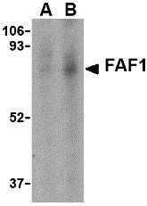
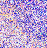
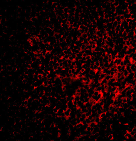
| Application Notes | FAF1 antibody can be used for detection of FAF1 by Western blot at 1 - 2 µg/mL. Antibody can also be used for immunohistochemistry starting at 2.5 µg/mL. For immunofluorescence start at 20 µg/mL. |
| Gene ID | 11124 |
|---|---|
| Other Names | FAF1 Antibody: hFAF1, CGI-03, HFAF1s, UBXD12, UBXN3A, FAS-associated factor 1, UBX domain-containing protein 12, hFAF1, Fas (TNFRSF6) associated factor 1 |
| Target/Specificity | FAF1; |
| Reconstitution & Storage | FAF1 antibody can be stored at 4℃ for three months and -20℃, stable for up to one year. As with all antibodies care should be taken to avoid repeated freeze thaw cycles. Antibodies should not be exposed to prolonged high temperatures. |
| Precautions | FAF1 Antibody is for research use only and not for use in diagnostic or therapeutic procedures. |
| Name | FAF1 |
|---|---|
| Synonyms | UBXD12, UBXN3A |
| Function | Ubiquitin-binding protein (PubMed:19722279). Required for the progression of DNA replication forks by targeting DNA replication licensing factor CDT1 for degradation (PubMed:26842564). Potentiates but cannot initiate FAS-induced apoptosis (By similarity). |
| Cellular Location | Nucleus. |
| Tissue Location | Most abundant in testis, slightly less abundant in skeletal muscle and heart, followed by prostate, thymus, ovary, small intestine, and colon. Not detected in the peripheral blood leukocytes |
For Research Use Only. Not For Use In Diagnostic Procedures.
Provided below are standard protocols that you may find useful for product applications.
BACKGROUND
FAF1 Antibody: Fas-associated protein 1 (FAF1) was initially identified as a Fas-binding pro-apoptotic protein that is component of the death-inducing signaling complex in Fas-mediated apoptosis. FAF1 can also induce apoptosis in the absence of extrinsic death signals when overexpressed although it does not contain typical death motifs such as the death domain, death effector domain, and caspase recruitment domain. Overexpression of FAF1 also decreases the basal level of NF-κB activity in transfected 293 cells, inhibits NF-κB activity induced by TNF-α, IL-1β and lipopolysaccharide, and prevents NF-κB translocation to the nucleus, suggesting that another role of FAF1 is to negatively regulate the activity of NF-κB. FAF1 can also interact with the inflammatory signaling PYRIN-containing Apaf-1-like proteins (PYPAFs, also called NALPs) such as PYPAF1, PYPAF2 (NALP2), and PYPAF7, suggesting FAF1 may also be involved in the inflammation pathway. Multiple differentially spliced isoforms of FAF1 are known to exist.
REFERENCES
Chu K, Niu X, and Williams LT. A Fas-associated protein factor, FAF1, potentiates Fas-mediated apoptosis. Proc. Natl. Acad. Sci. USA1995; 92:11894-8.
Ryu SW and Kim E. Apoptosis induced by human Fas-associated factor 1, hFAF1, requires its ubiquitin homologous domain, but not the Fas-binding domain. Biochem. Biophys. Res. Commun. 2001; 286:1027-32.
Park M-Y, Jang HD, Lee SY, et al. Fas-associated Factor-1 inhibits Nuclear Factor-κB (NF-κB) activity by interfering with nuclear translocation of the RelA (p65) subunit of NF-κB. J. Biol. Chem.2004; 279:2544-9.
Kinoshita T, Kondoh C, Hasegawa M, et al. Fas-associated factor 1 is a negative regulator of PYRIN-containing Apaf-1-like protein 1. Int. Immunol. 2006;18:1701-6.
终于等到您。ABCEPTA(百远生物)抗体产品。
点击下方“我要评价 ”按钮提交您的反馈信息,您的反馈和评价是我们最宝贵的财富之一,
我们将在1-3个工作日内处理您的反馈信息。
如有疑问,联系:0512-88856768 tech-china@abcepta.com.


