


 癌症的基本特征包括细胞增殖、血管生成、迁移、凋亡逃避机制和细胞永生等。找到癌症发生过程中这些通路的关键标记物和对应的抗体用于检测至关重要。
 为您推荐一个泛素化位点预测神器——泛素化分析工具,可以为您的蛋白的泛素化位点作出预测和评分。
 细胞自噬受体图形绘图工具为你的蛋白的细胞受体结合位点作出预测和评分,识别结合到自噬通路中的蛋白是非常重要的,便于让我们理解自噬在正常生理、病理过程中的作用,如发育、细胞分化、神经退化性疾病、压力条件下、感染和癌症。
VISA Antibody

- 产品详情
- 实验流程
- 背景知识
Application
| WB, IF, E, IHC-P |
|---|---|
| Primary Accession | Q7Z434 |
| Other Accession | NP_065797, 83776598 |
| Reactivity | Human, Mouse, Rat |
| Host | Rabbit |
| Clonality | Polyclonal |
| Isotype | IgG |
| Calculated MW | 56528 Da |
| Concentration (mg/ml) | 1 mg/mL |
| Conjugate | Unconjugated |
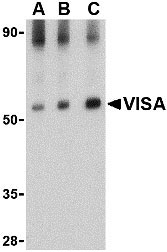
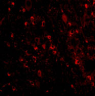
| Application Notes | VISA antibody can be used for detection of VISA by Western blot at 0.5 - 2 µg/mL. Antibody can also be used for immunohistochemistry starting at 2.5 µg/mL. For immunofluorescence start at 20 µg/mL. |
| Gene ID | 57506 |
|---|---|
| Other Names | VISA Antibody: IPS1, VISA, IPS-1, CARDIF, IPS1, KIAA1271, Mitochondrial antiviral-signaling protein, CARD adapter inducing interferon beta, MAVS, mitochondrial antiviral signaling protein |
| Target/Specificity | MAVS; |
| Reconstitution & Storage | VISA antibody can be stored at 4℃ for three months and -20℃, stable for up to one year. As with all antibodies care should be taken to avoid repeated freeze thaw cycles. Antibodies should not be exposed to prolonged high temperatures. |
| Precautions | VISA Antibody is for research use only and not for use in diagnostic or therapeutic procedures. |
| Name | MAVS {ECO:0000303|PubMed:16125763, ECO:0000312|HGNC:HGNC:29233} |
|---|---|
| Function | Adapter required for innate immune defense against viruses (PubMed:16125763, PubMed:16127453, PubMed:16153868, PubMed:16177806, PubMed:19631370, PubMed:20127681, PubMed:20451243, PubMed:21170385, PubMed:23087404, PubMed:27992402, PubMed:33139700, PubMed:37582970). Acts downstream of DHX33, RIGI and IFIH1/MDA5, which detect intracellular dsRNA produced during viral replication, to coordinate pathways leading to the activation of NF-kappa-B, IRF3 and IRF7, and to the subsequent induction of antiviral cytokines such as IFNB and RANTES (CCL5) (PubMed:16125763, PubMed:16127453, PubMed:16153868, PubMed:16177806, PubMed:19631370, PubMed:20127681, PubMed:20451243, PubMed:20628368, PubMed:21170385, PubMed:23087404, PubMed:25636800, PubMed:27736772, PubMed:33110251). Peroxisomal and mitochondrial MAVS act sequentially to create an antiviral cellular state (PubMed:20451243). Upon viral infection, peroxisomal MAVS induces the rapid interferon-independent expression of defense factors that provide short-term protection, whereas mitochondrial MAVS activates an interferon-dependent signaling pathway with delayed kinetics, which amplifies and stabilizes the antiviral response (PubMed:20451243). May activate the same pathways following detection of extracellular dsRNA by TLR3 (PubMed:16153868). May protect cells from apoptosis (PubMed:16125763). Involved in NLRP3 inflammasome activation by mediating NLRP3 recruitment to mitochondria (PubMed:23582325). |
| Cellular Location | Mitochondrion outer membrane; Single-pass membrane protein. Mitochondrion. Peroxisome |
| Tissue Location | Present in T-cells, monocytes, epithelial cells and hepatocytes (at protein level). Ubiquitously expressed, with highest levels in heart, skeletal muscle, liver, placenta and peripheral blood leukocytes. |
For Research Use Only. Not For Use In Diagnostic Procedures.
Provided below are standard protocols that you may find useful for product applications.
BACKGROUND
VISA Antibody: Two distinct signaling pathways activate the host innate immunity against viral infection. One pathway is reliant on members of the Toll-like receptor (TLR) family while the other uses the RNA helicase RIG-I as a receptor for intracellular viral double-stranded RNA as a trigger for the immune response. VISA is a mitochondrial membrane protein that was identified as a critical component in the IFN-b signaling pathways that recruits IRF-3 to RIG-I, leading to its activation and that of NF-κB. VISA is also thought to interact with other components of the innate immune pathway such as the TLR adapter protein TRIF, TRAF2 and TRAF6. VISA also interacts with the IKKα, IKKβ and IKKε kinases through its C-terminal region. Cleavage of this region by the Hepatitis C virus (HCV) protease allows HCV to escape the host immune system. At least three isoforms of VISA are known to exist.
REFERENCES
Seth RB, Sun L, and Chen ZJ. Antiviral innate immunity pathways. Cell Res.2006; 16:141-7.
Xu LG, Wang YY, Han KJ, et al. VISA is an adapter protein required for virus-triggered IFN-beta signaling. Mol. Cell2005; 19:727-40.
Meylan E, Curran J, Hofman K, et al. Cardif is an adaptor protein in the RIG-I antiviral pathway and is targeted by hepatitis C virus. Nature2005; 1167-72.
终于等到您。ABCEPTA(百远生物)抗体产品。
点击下方“我要评价 ”按钮提交您的反馈信息,您的反馈和评价是我们最宝贵的财富之一,
我们将在1-3个工作日内处理您的反馈信息。
如有疑问,联系:0512-88856768 tech-china@abcepta.com.


