


 癌症的基本特征包括细胞增殖、血管生成、迁移、凋亡逃避机制和细胞永生等。找到癌症发生过程中这些通路的关键标记物和对应的抗体用于检测至关重要。
 为您推荐一个泛素化位点预测神器——泛素化分析工具,可以为您的蛋白的泛素化位点作出预测和评分。
 细胞自噬受体图形绘图工具为你的蛋白的细胞受体结合位点作出预测和评分,识别结合到自噬通路中的蛋白是非常重要的,便于让我们理解自噬在正常生理、病理过程中的作用,如发育、细胞分化、神经退化性疾病、压力条件下、感染和癌症。
PDL-2 Antibody
- 产品详情
- 实验流程
- 背景知识
Application
| WB, IF, E, IHC-P |
|---|---|
| Primary Accession | Q9BQ51 |
| Other Accession | NP_079515, 190014605 |
| Reactivity | Human, Mouse, Rat |
| Host | Rabbit |
| Clonality | Polyclonal |
| Isotype | IgG |
| Calculated MW | 30957 Da |
| Concentration (mg/ml) | 1 mg/mL |
| Conjugate | Unconjugated |
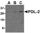
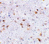
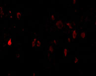
| Application Notes | PDL-2 antibody can be used for detection of PDL-2 by Western blot at 0.5 - 2 µg/mL. Antibody can also be used for immunohistochemistry starting at 2.5 µg/mL. For immunofluorescence start at 20 µg/mL. |
| Gene ID | 80380 |
|---|---|
| Other Names | PDL-2 Antibody: B7DC, Btdc, PDL2, CD273, PD-L2, PDCD1L2, bA574F11.2, B7DC, Programmed cell death 1 ligand 2, Butyrophilin B7-DC, PD-1 ligand 2, programmed cell death 1 ligand 2 |
| Target/Specificity | PDCD1LG2; |
| Reconstitution & Storage | PDL-2 antibody can be stored at 4℃ for three months and -20℃, stable for up to one year. As with all antibodies care should be taken to avoid repeated freeze thaw cycles. Antibodies should not be exposed to prolonged high temperatures. |
| Precautions | PDL-2 Antibody is for research use only and not for use in diagnostic or therapeutic procedures. |
| Name | PDCD1LG2 |
|---|---|
| Synonyms | B7DC, CD273, PDCD1L2, PDL2 |
| Function | Involved in the costimulatory signal, essential for T-cell proliferation and IFNG production in a PDCD1-independent manner. Interaction with PDCD1 inhibits T-cell proliferation by blocking cell cycle progression and cytokine production (By similarity). |
| Cellular Location | [Isoform 3]: Secreted [Isoform 1]: Cell membrane; Single-pass type I membrane protein {ECO:0000250|UniProtKB:Q9WUL5, ECO:0000305|PubMed:15340161} |
| Tissue Location | Highly expressed in heart, placenta, pancreas, lung and liver and weakly expressed in spleen, lymph nodes and thymus |
For Research Use Only. Not For Use In Diagnostic Procedures.
Provided below are standard protocols that you may find useful for product applications.
BACKGROUND
PDL-2 Antibody: Cell-mediated immune responses are initiated by T lymphocytes that are themselves stimulated by co gnate peptides bound to MHC molecules on antigen-presenting cells (APC). T-cell activation is generally self-limited as activated T cells express receptors such as PD-1 (also known as PDCD-1) that mediate inhibitory signals from the APC. PD-1 can bind two different but related ligands, PDL-1 and PDL-2, both of which are thought act as a negative regulator of T cell activation. However, it has been suggested that PDL-2 can act to stimulate an immunogenic response through and alternative receptor from PD-1.
REFERENCES
Holling TM, Schooten E, and van Den Elsing PJ. Function and regulation of MHC class II molecules in T-lymphocytes: of mice and men. Hum. Immunol. 2004; 65:282-90.
Ishida Y, Agata Y, Shibahara K, et al. Induced expression of PD-1, a novel member of the immunoglobulin gene superfamily, upon programmed cell death. EMBO J. 1992; 11:3887-95.
LaGier J and Pober JS. Immune accessory functions of human endothelial cells are modulated by overexpression of B7-H1 (PDL1). Hum. Immunol. 2006; 67:568-78.
Zhang Y, Chung Y, Bishop C, et al. Regulation of T cell activation and tolerance by PDL2. Proc. Natl. Acad. Sci. USA 2006; 103:11695-700.
终于等到您。ABCEPTA(百远生物)抗体产品。
点击下方“我要评价 ”按钮提交您的反馈信息,您的反馈和评价是我们最宝贵的财富之一,
我们将在1-3个工作日内处理您的反馈信息。
如有疑问,联系:0512-88856768 tech-china@abcepta.com.


