


 癌症的基本特征包括细胞增殖、血管生成、迁移、凋亡逃避机制和细胞永生等。找到癌症发生过程中这些通路的关键标记物和对应的抗体用于检测至关重要。
 为您推荐一个泛素化位点预测神器——泛素化分析工具,可以为您的蛋白的泛素化位点作出预测和评分。
 细胞自噬受体图形绘图工具为你的蛋白的细胞受体结合位点作出预测和评分,识别结合到自噬通路中的蛋白是非常重要的,便于让我们理解自噬在正常生理、病理过程中的作用,如发育、细胞分化、神经退化性疾病、压力条件下、感染和癌症。
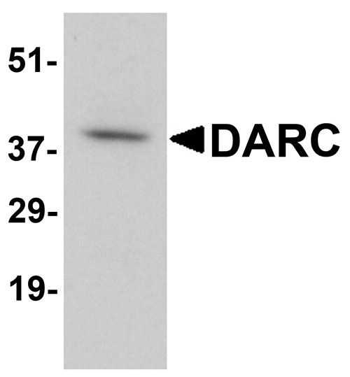
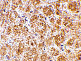
DARC Antibody
- 产品详情
- 实验流程
- 背景知识
Application 
| WB, E, IHC-P |
|---|---|
| Primary Accession | Q16570 |
| Other Accession | Q16570, 67476970 |
| Reactivity | Human, Mouse, Rat |
| Host | Rabbit |
| Clonality | Polyclonal |
| Isotype | IgG |
| Calculated MW | 35553 Da |
| Concentration (mg/ml) | 1 mg/mL |
| Conjugate | Unconjugated |
| Application Notes | DARC antibody can be used for detection of DARC by Western blot at 0.5 - 2 µg/mL. Antibody can also be used for immunohistochemistry starting at 2.5 µg/mL. |
| Gene ID | 2532 |
|---|---|
| Other Names | DARC Antibody: FY, Dfy, GPD, GpFy, ACKR1, CCBP1, CD234, WBCQ1, FY, Atypical chemokine receptor 1, Duffy blood group, chemokine receptor |
| Target/Specificity | DARC; |
| Reconstitution & Storage | DARC antibody can be stored at 4℃ for three months and -20℃, stable for up to one year. As with all antibodies care should be taken to avoid repeated freeze thaw cycles. Antibodies should not be exposed to prolonged high temperatures. |
| Precautions | DARC Antibody is for research use only and not for use in diagnostic or therapeutic procedures. |
For Research Use Only. Not For Use In Diagnostic Procedures.
| Name | ACKR1 |
|---|---|
| Function | Atypical chemokine receptor that controls chemokine levels and localization via high-affinity chemokine binding that is uncoupled from classic ligand-driven signal transduction cascades, resulting instead in chemokine sequestration, degradation, or transcytosis. Also known as interceptor (internalizing receptor) or chemokine-scavenging receptor or chemokine decoy receptor. Has a promiscuous chemokine- binding profile, interacting with inflammatory chemokines of both the CXC and the CC subfamilies but not with homeostatic chemokines. Acts as a receptor for chemokines including CCL2, CCL5, CCL7, CCL11, CCL13, CCL14, CCL17, CXCL5, CXCL6, IL8/CXCL8, CXCL11, GRO, RANTES, MCP-1 and TARC. May regulate chemokine bioavailability and, consequently, leukocyte recruitment through two distinct mechanisms: when expressed in endothelial cells, it sustains the abluminal to luminal transcytosis of tissue-derived chemokines and their subsequent presentation to circulating leukocytes; when expressed in erythrocytes, serves as blood reservoir of cognate chemokines but also as a chemokine sink, buffering potential surges in plasma chemokine levels. (Microbial infection) Acts as a receptor for the malaria parasite Plasmodium knowlesi. |
| Cellular Location | Early endosome. Recycling endosome. Membrane; Multi-pass membrane protein. Note=Predominantly localizes to endocytic vesicles, and upon stimulation by the ligand is internalized via caveolae. Once internalized, the ligand dissociates from the receptor, and is targeted to degradation while the receptor is recycled back to the cell membrane |
| Tissue Location | Found in adult kidney, adult spleen, bone marrow and fetal liver. In particular, it is expressed along postcapillary venules throughout the body, except in the adult liver. Erythroid cells and postcapillary venule endothelium are the principle tissues expressing duffy. Fy(-A-B) individuals do not express duffy in the bone marrow, however they do, in postcapillary venule endothelium |
Provided below are standard protocols that you may find useful for product applications.
BACKGROUND
DARC Antibody: DARC, also known as the Duffy antigen/chemokine receptor, is a seven-transmembrane protein homologous to the classical chemokine G-protein coupled receptors (GPCRs) with the exception of the motif required for G protein coupling. DARC can bind with high affinity several chemokines without transducing any signal, suggesting it may modulate the signals normally induced by these chemokines. Recently, DARC was found to interact with KAI1, a four transmembrane protein recently identified as a tumor metastasis suppressor protein. It is thought that tumor cells dislodged from the primary tumor and expressing KAI1 interact with DARC proteins expressed on vascular cells, transmitting a senescent signal to the tumor cells, while tumor cells that have lost KAI1 expression can proliferate and potentially give rise to metastases. At least three isoforms of DARC are known to exist.
REFERENCES
Chaudhuri A, Polyakova J, Zbrzezna V, et al. Cloning of glycoprotein D cDNA, which encodes the major subunit of the Duffy blood group system and the receptor for the Plasmodium vivax malaria parasite. Proc. Natl. Acad. Sci. USA1993; 90:10793-7.
Gardner L, Patterson AM, Ashton BA, et al. The human Duffy antigen binds selected inflammatory but not homeostatic chemokines. Biochem. Biophys. Res. Commun.2004; 321:306-12.
Gil ML, Vita N, Lebel-Binay S, et al. A member of the tetra spans transmembrane protein superfamily is recognized by a monoclonal antibody raised against an HLA class I-deficient, lymphokine-activated killer-susceptible, B lymphocyte line. Cloning and functional studies. J. Immunol.1992; 2826-33.

终于等到您。ABCEPTA(百远生物)抗体产品。
点击下方“我要评价 ”按钮提交您的反馈信息,您的反馈和评价是我们最宝贵的财富之一,
我们将在1-3个工作日内处理您的反馈信息。
如有疑问,联系:0512-88856768 tech-china@abcepta.com.


