


 癌症的基本特征包括细胞增殖、血管生成、迁移、凋亡逃避机制和细胞永生等。找到癌症发生过程中这些通路的关键标记物和对应的抗体用于检测至关重要。
 为您推荐一个泛素化位点预测神器——泛素化分析工具,可以为您的蛋白的泛素化位点作出预测和评分。
 细胞自噬受体图形绘图工具为你的蛋白的细胞受体结合位点作出预测和评分,识别结合到自噬通路中的蛋白是非常重要的,便于让我们理解自噬在正常生理、病理过程中的作用,如发育、细胞分化、神经退化性疾病、压力条件下、感染和癌症。
KAI1 Antibody
- 产品详情
- 实验流程
- 背景知识
Application
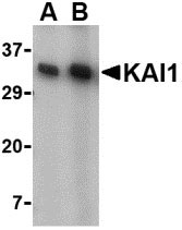
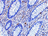
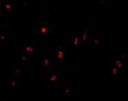
| WB, IF, E, IHC-P |
|---|---|
| Primary Accession | P27701 |
| Other Accession | NP_002222, 4504813 |
| Reactivity | Human, Mouse, Rat |
| Host | Rabbit |
| Clonality | Polyclonal |
| Isotype | IgG |
| Calculated MW | 29626 Da |
| Concentration (mg/ml) | 1 mg/mL |
| Conjugate | Unconjugated |
| Application Notes | KAI1 antibody can be used for detection of KAI1 by Western blot at 0.5 - 1 µg/mL. Antibody can also be used for immunohistochemistry starting at 2.5 µg/mL. For immunofluorescence start at 20 µg/mL. |
| Gene ID | 3732 |
|---|---|
| Other Names | KAI1 Antibody: R2, 4F9, C33, IA4, ST6, GR15, KAI1, SAR2, TSPAN27, CD82 antigen, C33 antigen, Tspan-27, CD82 molecule |
| Target/Specificity | CD82; |
| Reconstitution & Storage | KAI1 antibody can be stored at 4℃ for three months and -20℃, stable for up to one year. As with all antibodies care should be taken to avoid repeated freeze thaw cycles. Antibodies should not be exposed to prolonged high temperatures. |
| Precautions | KAI1 Antibody is for research use only and not for use in diagnostic or therapeutic procedures. |
| Name | CD82 |
|---|---|
| Synonyms | KAI1, SAR2, ST6, TSPAN27 |
| Function | Structural component of specialized membrane microdomains known as tetraspanin-enriched microdomains (TERMs), which act as platforms for receptor clustering and signaling (PubMed:19497983). Participates thereby in diverse biological functions such as cell signal transduction, adhesion, migration and protein trafficking. Acts as a attenuator of EGF signaling, facilitating ligand-induced endocytosis of the receptor and its subsequent desensitization (PubMed:10985391, PubMed:35538033). Mechanistically, modulates ligand- induced ubiquitination and trafficking of EGFR via E3 ligase CBL phosphorylation by PKC (PubMed:23897813). Increases cell-matrix adhesion by regulating the membrane organization of integrin alpha4/ITA4 (PubMed:24623721, PubMed:8757325). Modulates adhesion and suppresses cell migration through other integrins such as the alpha6/ITGA6 and beta1/ITGB1 (PubMed:15557282, PubMed:17560548). Decreases cell-associated plasminogen activation by interfering with the interaction between urokinase-type plasminogen activator/PLAU and its receptor PLAUR (PubMed:15677461). Associates with CD4 or CD8 and delivers costimulatory signals for the TCR/CD3 pathway. Plays a role in TLR9 trafficking to acidified CpG-containing compartments by controlling interaction between TLR9 and VAMP3 and subsequent myddosome assembly (By similarity). Inhibits LPS-induced inflammatory response by preventing binding of LPS to TLR4 on the cell surface (PubMed:36945827). Plays a role in the activation of macrophages into anti-inflammatory phenotypes (By similarity). Independently of Toll- like receptor (TLR) signaling, is recruited to pathogen-containing phagosomes prior to fusion with lysosomes and thereby participates in antigen presentation (By similarity). Also acts to control angiogenesis and switch angiogenic milieu to quiescent state by binding and sequestering VEGFA and PDGFB to inhibit the signaling they trigger via their respective cell surface receptor (PubMed:34530889). |
| Cellular Location | Cell membrane {ECO:0000269|PubMed:19497983, ECO:0000269|PubMed:23897813, ECO:0000269|PubMed:30463011, ECO:0000269|PubMed:34530889, ECO:0000269|PubMed:8757325, ECO:0000269|Ref.4}; Multi-pass membrane protein Cytoplasmic vesicle, phagosome {ECO:0000250|UniProtKB:P40237} |
| Tissue Location | Lymphoid specific. |
For Research Use Only. Not For Use In Diagnostic Procedures.
Provided below are standard protocols that you may find useful for product applications.
BACKGROUND
KAI1 Antibody: KAI1 was initially identified from a T-cell activation study as a four-transmembrane protein that plays an accessory role in T-cell activation, and was later determined to act as a cancer metastasis suppressor gene. This protein is ubiquitously expressed at moderate to high levels in most tissues, but its expression is downregulated during tumor progression. The loss of KAI1 and p53 is associated with poor survival for prostate and other cancer patients. Recently, KAI1 was found to interact with DARC, the Duffy antigen for chemokines using a yeast two hybrid screen. It is thought that tumor cells dislodged from the primary tumor and expressing KAI1 interact with DARC proteins expressed on vascular cells, transmitting a senescent signal to the tumor cells, while tumor cells that have lost KAI1 expression can proliferate and potentially give rise to metastases. At least two isoforms of KAI1 are known to exist.
REFERENCES
HW Gaugitsch, Hofer E, Huber NE, et al. A new superfamily of lymphoid and melanoma cell proteins with extensive homology to Schistosoma mansoni antigen SM23. Eur. J. Immunol.1991; 21:377-83.
Gil ML, Vita N, Lebel-Binay S, et al. A member of the tetra spans transmembrane protein superfamily is recognized by a monoclonal antibody raised against an HLA class I-deficient, lymphokine-activated killer-susceptible, B lymphocyte line. Cloning and functional studies. J. Immunol.1992; 2826-33.
Dong JT, Lamb PW, Rinker-Schaeffer CW, et al. KAI1, a metastasis suppressor gene for prostate cancer on human chromosome 11p11.2. Science1995; 884-86.
Kauffman EC, Robinson VL, Stadler WM, et al. Metastasis suppression: the evolving role of metastasis suppressor genes for regulating cancer cell growth at the secondary site. J. Urol.2003; 169:1122-33.
终于等到您。ABCEPTA(百远生物)抗体产品。
点击下方“我要评价 ”按钮提交您的反馈信息,您的反馈和评价是我们最宝贵的财富之一,
我们将在1-3个工作日内处理您的反馈信息。
如有疑问,联系:0512-88856768 tech-china@abcepta.com.


