


 癌症的基本特征包括细胞增殖、血管生成、迁移、凋亡逃避机制和细胞永生等。找到癌症发生过程中这些通路的关键标记物和对应的抗体用于检测至关重要。
 为您推荐一个泛素化位点预测神器——泛素化分析工具,可以为您的蛋白的泛素化位点作出预测和评分。
 细胞自噬受体图形绘图工具为你的蛋白的细胞受体结合位点作出预测和评分,识别结合到自噬通路中的蛋白是非常重要的,便于让我们理解自噬在正常生理、病理过程中的作用,如发育、细胞分化、神经退化性疾病、压力条件下、感染和癌症。
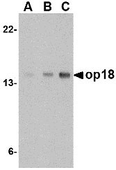
op18 Antibody
- 产品详情
- 实验流程
- 背景知识
Application
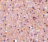
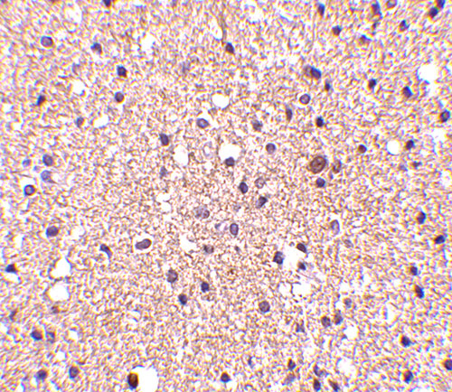
| WB, IF, E, IHC-P |
|---|---|
| Primary Accession | P16949 |
| Other Accession | AAH82228, 51895905 |
| Reactivity | Human, Mouse |
| Host | Rabbit |
| Clonality | Polyclonal |
| Isotype | IgG |
| Calculated MW | 17303 Da |
| Concentration (mg/ml) | 1 mg/mL |
| Conjugate | Unconjugated |
| Application Notes | Op18 antibody can be used for detection of op18 by Western blot at 0.5 - 2 µg/mL. Antibody can also be used for immunohistochemistry starting at 2.5 µg/mL. For immunofluorescence start at 20 µg/mL. |
| Gene ID | 3925 |
|---|---|
| Other Names | Stathmin, Leukemia-associated phosphoprotein p18, Metablastin, Oncoprotein 18, Op18, Phosphoprotein p19, pp19, Prosolin, Protein Pr22, pp17, STMN1, C1orf215, LAP18, OP18 |
| Target/Specificity | STMN1; |
| Reconstitution & Storage | op18 antibody can be stored at 4℃ for three months and -20℃, stable for up to one year. As with all antibodies care should be taken to avoid repeated freeze thaw cycles. Antibodies should not be exposed to prolonged high temperatures. |
| Precautions | op18 Antibody is for research use only and not for use in diagnostic or therapeutic procedures. |
| Name | STMN1 |
|---|---|
| Synonyms | C1orf215, LAP18, OP18 |
| Function | Involved in the regulation of the microtubule (MT) filament system by destabilizing microtubules. Prevents assembly and promotes disassembly of microtubules. Phosphorylation at Ser-16 may be required for axon formation during neurogenesis. Involved in the control of the learned and innate fear (By similarity). |
| Cellular Location | Cytoplasm, cytoskeleton. |
| Tissue Location | Ubiquitous. Expression is strongest in fetal and adult brain, spinal cord, and cerebellum, followed by thymus, bone marrow, testis, and fetal liver. Expression is intermediate in colon, ovary, placenta, uterus, and trachea, and is readily detected at substantially lower levels in all other tissues examined. Lowest expression is found in adult liver. Present in much greater abundance in cells from patients with acute leukemia of different subtypes than in normal peripheral blood lymphocytes, non-leukemic proliferating lymphoid cells, bone marrow cells, or cells from patients with chronic lymphoid or myeloid leukemia. |
For Research Use Only. Not For Use In Diagnostic Procedures.
Provided below are standard protocols that you may find useful for product applications.
BACKGROUND
op18 Antibody: Op18 belongs to the stathmin family of genes and encodes a ubiquitous cytosolic phosphoprotein that may function as an intracellular relay integrating several signaling pathways such as those involved in cell proliferation and differentiation. Op18 has also been shown to be involved in the regulation of the microtubule filament system by destabilizing microtubules, thereby preventing assembly and promoting the disassembly of microtubules. More recently, op18 has been implicated as a potential target of the ASK1-p38 MAP kinase cascade, suggesting that the ASK1-p38 cascade may regulate microtubule dynamics through op18. Op18 is highly expressed in a wide variety of human malignancies, including leukemia, prostate cancer, ovarian carcinoma, and breast carcinoma, suggesting that op18 may be an ideal target for anti-cancer therapeutics.
REFERENCES
Curmi PA, Gavet O, Charbaut E, et al. Stathmin and its phosphoprotein family: general properties, biochemical and functional interaction with tubulin. Cell Structure and Function 1999; 24:345-57.
Belmont LD and Mitchison TJ. Identification of a protein that interacts with tubulin dimers and increases catastrophe rate of microtubules. Cell 1996; 84:623-31.
Mizumura K, Takeda K, Hashimoto S, et al. Identification of op18/stathmin as a potential target of ASK1-p38 MAP kinase cascade. J. Cell Physiol. 2006; 206:363-70.
Mistry SJ and Atweh GF. Role of stathmin in the regulation of the mitotic spindle. Mount Sinai J. Med. 2002; 69:299-304.
终于等到您。ABCEPTA(百远生物)抗体产品。
点击下方“我要评价 ”按钮提交您的反馈信息,您的反馈和评价是我们最宝贵的财富之一,
我们将在1-3个工作日内处理您的反馈信息。
如有疑问,联系:0512-88856768 tech-china@abcepta.com.


