


 癌症的基本特征包括细胞增殖、血管生成、迁移、凋亡逃避机制和细胞永生等。找到癌症发生过程中这些通路的关键标记物和对应的抗体用于检测至关重要。
 为您推荐一个泛素化位点预测神器——泛素化分析工具,可以为您的蛋白的泛素化位点作出预测和评分。
 细胞自噬受体图形绘图工具为你的蛋白的细胞受体结合位点作出预测和评分,识别结合到自噬通路中的蛋白是非常重要的,便于让我们理解自噬在正常生理、病理过程中的作用,如发育、细胞分化、神经退化性疾病、压力条件下、感染和癌症。
LIS1 Antibody
- 产品详情
- 实验流程
- 背景知识
Application
| WB, IF, ICC, E |
|---|---|
| Primary Accession | P43034 |
| Other Accession | P43034, 1170794 |
| Reactivity | Human, Mouse, Rat |
| Host | Rabbit |
| Clonality | Polyclonal |
| Isotype | IgG |
| Calculated MW | 46638 Da |
| Concentration (mg/ml) | 1 mg/mL |
| Conjugate | Unconjugated |
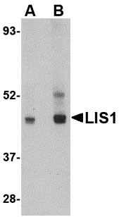
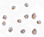
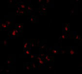
| Application Notes | LIS1 antibody can be used for detection of LIS1 by Western blot at 0.5 - 1 µg/mL. Antibody can also be used for immunocytochemistry starting at 2.5 µg/mL. For immunofluorescence start at 20 µg/mL. |
| Gene ID | 5048 |
|---|---|
| Other Names | Platelet-activating factor acetylhydrolase IB subunit alpha {ECO:0000255|HAMAP-Rule:MF_03141}, Lissencephaly-1 protein {ECO:0000255|HAMAP-Rule:MF_03141}, LIS-1 {ECO:0000255|HAMAP-Rule:MF_03141}, PAF acetylhydrolase 45 kDa subunit {ECO:0000255|HAMAP-Rule:MF_03141}, PAF-AH 45 kDa subunit {ECO:0000255|HAMAP-Rule:MF_03141}, PAF-AH alpha {ECO:0000255|HAMAP-Rule:MF_03141}, PAFAH alpha {ECO:0000255|HAMAP-Rule:MF_03141}, PAFAH1B1 {ECO:0000255|HAMAP-Rule:MF_03141} |
| Target/Specificity | PAFAH1B1; |
| Reconstitution & Storage | LIS1 antibody can be stored at 4℃ for three months and -20℃, stable for up to one year. As with all antibodies care should be taken to avoid repeated freeze thaw cycles. Antibodies should not be exposed to prolonged high temperatures. |
| Precautions | LIS1 Antibody is for research use only and not for use in diagnostic or therapeutic procedures. |
| Name | LIS1 |
|---|---|
| Function | Regulatory subunit (beta subunit) of the cytosolic type I platelet-activating factor (PAF) acetylhydrolase (PAF-AH (I)), an enzyme that catalyzes the hydrolyze of the acetyl group at the sn-2 position of PAF and its analogs and participates in PAF inactivation. Regulates the PAF-AH (I) activity in a catalytic dimer composition- dependent manner (By similarity). Required for proper activation of Rho GTPases and actin polymerization at the leading edge of locomoting cerebellar neurons and postmigratory hippocampal neurons in response to calcium influx triggered via NMDA receptors (By similarity). Positively regulates the activity of the minus-end directed microtubule motor protein dynein. May enhance dynein-mediated microtubule sliding by targeting dynein to the microtubule plus end. Required for several dynein- and microtubule-dependent processes such as the maintenance of Golgi integrity, the peripheral transport of microtubule fragments and the coupling of the nucleus and centrosome. Required during brain development for the proliferation of neuronal precursors and the migration of newly formed neurons from the ventricular/subventricular zone toward the cortical plate. Neuronal migration involves a process called nucleokinesis, whereby migrating cells extend an anterior process into which the nucleus subsequently translocates. During nucleokinesis dynein at the nuclear surface may translocate the nucleus towards the centrosome by exerting force on centrosomal microtubules. May also play a role in other forms of cell locomotion including the migration of fibroblasts during wound healing. Required for dynein recruitment to microtubule plus ends and BICD2-bound cargos (PubMed:22956769). May modulate the Reelin pathway through interaction of the PAF-AH (I) catalytic dimer with VLDLR (By similarity). |
| Cellular Location | Cytoplasm, cytoskeleton. Cytoplasm, cytoskeleton, microtubule organizing center, centrosome. Cytoplasm, cytoskeleton, spindle {ECO:0000255|HAMAP-Rule:MF_03141}. Nucleus membrane {ECO:0000255|HAMAP- Rule:MF_03141}. Note=Redistributes to axons during neuronal development. Also localizes to the microtubules of the manchette in elongating spermatids and to the meiotic spindle in spermatocytes (By similarity). Localizes to the plus end of microtubules and to the centrosome. May localize to the nuclear membrane. |
| Tissue Location | Fairly ubiquitous expression in both the frontal and occipital areas of the brain |
For Research Use Only. Not For Use In Diagnostic Procedures.
Provided below are standard protocols that you may find useful for product applications.
BACKGROUND
LIS1 Antibody: Lissencephaly is a severe brain developmental disease characterized by the mislocalization of cortical neurons, a smooth cerebral surface, mental retardation, and seizures. Classical lissencephaly is caused by sporadic mutations in the LIS1 gene. While LIS1 is known to act in a pathway deactivating the lipid messenger platelet-activating factor, LIS1 forms a complex with Nudel and 14-3-3epsilon which is then transported from neuronal cell bodies through the actions of DISC1 and KIF5A, a microtubule-dependent directed motor protein kinesin. Decreased expression of LIS1 blocked neural stem cell division, morphogenesis, and motility, suggesting that LIS1 plays an important role in neuronal cell proliferation and localization in the developing brain. At least two isoforms of LIS1 are known to exist.
REFERENCES
McManus MF and Golden JA. Neuronal migration in developmental disorders. J. Child Neurol.2005; 20:280-6.
Reiner O, Carrozzo R, Shen Y, et al. Isolation of a Miller-Dieker lissencephaly gene containing G protein b-subunit-like repeats. Nature1993; 364:717-21.
Hattori M, Adachi H, Tsujimoto M, et al. Miller-Dieker lissencephaly gene encodes a subunit of brain platelet activating factor. Nature1994; 370:216-8.
Taya S, Shinoda T, Tsuboi D, et al. DISC1 regulates the transport of the NUDEL/LIS1/14-3-3e complex through kinesin-1. J. Neurosci.2007; 27:15-26.
终于等到您。ABCEPTA(百远生物)抗体产品。
点击下方“我要评价 ”按钮提交您的反馈信息,您的反馈和评价是我们最宝贵的财富之一,
我们将在1-3个工作日内处理您的反馈信息。
如有疑问,联系:0512-88856768 tech-china@abcepta.com.


