


 癌症的基本特征包括细胞增殖、血管生成、迁移、凋亡逃避机制和细胞永生等。找到癌症发生过程中这些通路的关键标记物和对应的抗体用于检测至关重要。
 为您推荐一个泛素化位点预测神器——泛素化分析工具,可以为您的蛋白的泛素化位点作出预测和评分。
 细胞自噬受体图形绘图工具为你的蛋白的细胞受体结合位点作出预测和评分,识别结合到自噬通路中的蛋白是非常重要的,便于让我们理解自噬在正常生理、病理过程中的作用,如发育、细胞分化、神经退化性疾病、压力条件下、感染和癌症。
ZBP1 Antibody
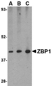
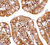
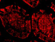
- 产品详情
- 实验流程
- 背景知识
Application 
| WB, IF, E, IHC-P |
|---|---|
| Primary Accession | Q9H171 |
| Other Accession | EAW75510, 119595916 |
| Reactivity | Human, Mouse, Rat |
| Host | Rabbit |
| Clonality | Polyclonal |
| Isotype | IgG |
| Calculated MW | 46343 Da |
| Concentration (mg/ml) | 1 mg/mL |
| Conjugate | Unconjugated |
| Application Notes | ZBP1 antibody can be used for the detection of ZBP1 by Western blot at 0.5 - 2 µg/mL. Antibody can also be used for immunohistochemistry starting at 2.5 µg/mL. For immunofluorescence start at 20 µg/mL. |
| Gene ID | 81030 |
|---|---|
| Other Names | Z-DNA-binding protein 1, Tumor stroma and activated macrophage protein DLM-1, ZBP1, C20orf183, DLM1 |
| Target/Specificity | ZBP1; Multiple isoforms of ZBP1 are known to exist. |
| Reconstitution & Storage | ZBP1 antibody can be stored at 4℃ for three months and -20℃, stable for up to one year. As with all antibodies care should be taken to avoid repeated freeze thaw cycles. Antibodies should not be exposed to prolonged high temperatures. |
| Precautions | ZBP1 Antibody is for research use only and not for use in diagnostic or therapeutic procedures. |
For Research Use Only. Not For Use In Diagnostic Procedures.
| Name | ZBP1 {ECO:0000303|PubMed:16876127, ECO:0000312|HGNC:HGNC:16176} |
|---|---|
| Function | Key innate sensor that recognizes and binds Z-RNA structures, which are produced by a number of viruses, such as herpesvirus, orthomyxovirus or flavivirus, and triggers different forms of cell death (PubMed:32200799). ZBP1 acts as an essential mediator of pyroptosis, necroptosis and apoptosis (PANoptosis), an integral part of host defense against pathogens, by activating RIPK3, caspase-8 (CASP8), and the NLRP3 inflammasome (By similarity). Key activator of necroptosis, a programmed cell death process in response to death- inducing TNF family members, via its ability to bind Z-RNA: once activated upon Z-RNA-binding, ZBP1 interacts and stimulates RIPK3 kinase, which phosphorylates and activates MLKL, triggering execution of programmed necrosis (By similarity). In addition to TNF-induced necroptosis, necroptosis can also take place in the nucleus in response to orthomyxoviruses infection: ZBP1 recognizes and binds Z-RNA structures that are produced in infected nuclei by orthomyxoviruses, such as the influenza A virus (IAV), leading to ZBP1 activation, RIPK3 stimulation and subsequent MLKL phosphorylation, triggering disruption of the nuclear envelope and leakage of cellular DNA into the cytosol (PubMed:32200799). ZBP1-dependent cell death in response to IAV infection promotes interleukin-1 alpha (IL1A) induction in an NLRP3- inflammasome-independent manner: IL1A expression is required for the optimal interleukin-1 beta (IL1B) production, and together, these cytokines promote infiltration of inflammatory neutrophils to the lung, leading to the formation of neutrophil extracellular traps (By similarity). In addition to its direct role in driving necroptosis via its ability to sense Z-RNAs, also involved in PANoptosis triggered in response to bacterial infection: component of the AIM2 PANoptosome complex, a multiprotein complex that triggers PANoptosis (By similarity). Also acts as the apical sensor of fungal infection responsible for activating PANoptosis (By similarity). Involved in CASP8-mediated cell death via its interaction with RIPK1 but independently of its ability to sense Z-RNAs (By similarity). In some cell types, also able to restrict viral replication by promoting cell death-independent responses (By similarity). In response to Zika virus infection in neurons, promotes a cell death-independent pathway that restricts viral replication: together with RIPK3, promotes a death- independent transcriptional program that modifies the cellular metabolism via up-regulation expression of the enzyme ACOD1/IRG1 and production of the metabolite itaconate (By similarity). Itaconate inhibits the activity of succinate dehydrogenase, generating a metabolic state in neurons that suppresses replication of viral genomes (By similarity). |
| Cellular Location | Cytoplasm. Nucleus. Note=Mainly cytoplasmic (PubMed:16876127, PubMed:16990255). Accumulates in the nucleus in response to influenza A virus (IAV) infection: senses IAV defective viral genomes RNA in the nucleus (By similarity). {ECO:0000250|UniProtKB:Q9QY24, ECO:0000269|PubMed:16876127, ECO:0000269|PubMed:16990255} |
| Tissue Location | Highly expressed in lymphatic tissues including lymph node, leukocytes, tonsil, bone marrow and spleen (PubMed:11842111). Expressed to a lesser extent in thymus, lung and liver (PubMed:11842111). |
Provided below are standard protocols that you may find useful for product applications.
BACKGROUND
ZBP1 Antibody: Z-DNA binding protein 1 (ZBP1) belongs to a family of proteins that contain the Zalpha domain which binds specifically to left-handed Z-DNA and Z-RNA. ZBP1 was initially identified as a novel gene that was up-regulated in activated macrophages in mice bearing ascites tumors, suggesting that is may play a role in processes such as host response in neoplasia. More recent reports indicate that the cytosolic ZBP1 has can act like the toll-like receptor TLR9 by detecting cytosolic double-stranded (ds) DNA and trigger induction of type I interferon and other innate immune responses. It is thought that the binding of ZBP1 to dsDNA enhances its association with innate immune response proteins such as the IRF3 transcription factor and the serine/threonine kinase TBK1 (also known as NAK).
REFERENCES
Schwartz T, Behlke J, Lowenhaupt K, et al. Structure of the DLM-1-Z-DNA complex reveals a conserved family of Z-DNA-binding proteins. Nat. Struct. Biol. 2001; 8:761-5.
Fu Y, Comella N, Tognazzi K, et al. Cloning of DLM-1, a novel gene that is up-regulated in active macrophages, using RNA differential display. Gene 1999; 204:157-63.
Takaoda A, Wang Z, Choi MK, et al. DAI (DLM-1/ZBP1) is a cytosolic DNA sensor and an activator of innate immune response. Nature 2007; 448:501-5.

终于等到您。ABCEPTA(百远生物)抗体产品。
点击下方“我要评价 ”按钮提交您的反馈信息,您的反馈和评价是我们最宝贵的财富之一,
我们将在1-3个工作日内处理您的反馈信息。
如有疑问,联系:0512-88856768 tech-china@abcepta.com.


