


 癌症的基本特征包括细胞增殖、血管生成、迁移、凋亡逃避机制和细胞永生等。找到癌症发生过程中这些通路的关键标记物和对应的抗体用于检测至关重要。
 为您推荐一个泛素化位点预测神器——泛素化分析工具,可以为您的蛋白的泛素化位点作出预测和评分。
 细胞自噬受体图形绘图工具为你的蛋白的细胞受体结合位点作出预测和评分,识别结合到自噬通路中的蛋白是非常重要的,便于让我们理解自噬在正常生理、病理过程中的作用,如发育、细胞分化、神经退化性疾病、压力条件下、感染和癌症。
BAP3 Antibody
- 产品详情
- 实验流程
- 背景知识
Application 
| WB, IF, E, IHC-P |
|---|---|
| Primary Accession | O94812 |
| Other Accession | BAA34710, 3929917 |
| Reactivity | Human |
| Host | Rabbit |
| Clonality | Polyclonal |
| Isotype | IgG |
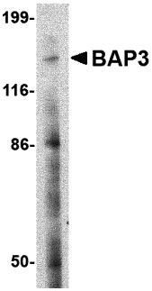
| Calculated MW | 131901 Da |
| Concentration (mg/ml) | 1 mg/mL |
| Conjugate | Unconjugated |
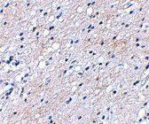
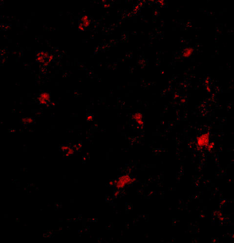
| Application Notes | BAP3 antibody can be used for the detection of BAP3 by Western blot at 2 µg/mL. Antibody can also be used for immunohistochemistry starting at 2.5 µg/mL. For immunofluorescence start at 20 µg/mL. |
| Gene ID | 8938 |
|---|---|
| Other Names | BAI1-associated protein 3, BAP3, BAIAP3, KIAA0734 |
| Target/Specificity | BAIAP3; |
| Reconstitution & Storage | BAP3 antibody can be stored at 4℃ for three months and -20℃, stable for up to one year. As with all antibodies care should be taken to avoid repeated freeze thaw cycles. Antibodies should not be exposed to prolonged high temperatures. |
| Precautions | BAP3 Antibody is for research use only and not for use in diagnostic or therapeutic procedures. |
For Research Use Only. Not For Use In Diagnostic Procedures.
| Name | BAIAP3 {ECO:0000303|PubMed:28626000, ECO:0000312|HGNC:HGNC:948} |
|---|---|
| Function | Functions in endosome to Golgi retrograde transport. In response to calcium influx, may interact with SNARE fusion receptors and membrane phospholipids to mediate endosome fusion with the trans- Golgi network. By promoting the recycling of secretory vesicle transmembrane proteins, it indirectly controls dense-core secretory vesicle biogenesis, maturation and their ability to mediate the constitutive and regulated secretion of neurotransmitters and hormones. May regulate behavior and food intake by controlling calcium-stimulated exocytosis of neurotransmitters including NPY and serotonin and hormones like insulin (PubMed:28626000). Proposed to play a role in hypothalamic neuronal firing by modulating gamma-aminobutyric acid (GABA)ergic inhibitory neurotransmission (By similarity). |
| Cellular Location | Cytoplasm, cytosol. Recycling endosome membrane; Peripheral membrane protein. Late endosome membrane; Peripheral membrane protein. Golgi apparatus, trans-Golgi network membrane; Peripheral membrane protein. Cell membrane; Peripheral membrane protein. Note=Rapidly recruited to the plasma membrane and to Golgi structures in response to increased intracellular calcium concentration. |
| Tissue Location | Predominantly expressed in brain (PubMed:9790924). Also expressed in nonneural tissues such as breast and testes epithelium (PubMed:12498718). |
Provided below are standard protocols that you may find useful for product applications.
BACKGROUND
BAP3 Antibody: BRAL1 is a member a superfamily consisting of several highly homologous hyaluronan and proteoglycan binding link proteins. BRAL1 is predominantly expressed in brain tissue and spinal cord. Like other members in the link-module superfamily, BRAL1 contains an immunoglobulin-like fold and two proteoglycan tandem repeats (PTRs). Its mRNA expression pattern is similar to other lectican proteoglycans, suggesting that BRAL1 may act to stabilize the binding between the extracellular matrix molecule hyaluronan and brevican. Immunostaining of mouse brain showed BRAL1 expression at P20 in the white matter of the developing cerebellum and in myelinated fiber tracts in the adult brain, suggesting that expression starts when axonal myelination occurs.
REFERENCES
Shiratsuchi T, Oda K, Nishimori H, et al. Cloning and characterization of BAP (BAI-associated protein 3), a C2 domain-containing protein that interacts with BAI1. Biochem. Biophys. Res. Comm.1998; 251:158-65.
Palmer RE, Lee SB, Wong JC, et al. Induction of BAIAP3 by the EWS-WT1 chimeric fusion implicates regulated exocytosis in tumorigenesis. Cancer Cell2002; 2:497-505.

终于等到您。ABCEPTA(百远生物)抗体产品。
点击下方“我要评价 ”按钮提交您的反馈信息,您的反馈和评价是我们最宝贵的财富之一,
我们将在1-3个工作日内处理您的反馈信息。
如有疑问,联系:0512-88856768 tech-china@abcepta.com.


