


 癌症的基本特征包括细胞增殖、血管生成、迁移、凋亡逃避机制和细胞永生等。找到癌症发生过程中这些通路的关键标记物和对应的抗体用于检测至关重要。
 为您推荐一个泛素化位点预测神器——泛素化分析工具,可以为您的蛋白的泛素化位点作出预测和评分。
 细胞自噬受体图形绘图工具为你的蛋白的细胞受体结合位点作出预测和评分,识别结合到自噬通路中的蛋白是非常重要的,便于让我们理解自噬在正常生理、病理过程中的作用,如发育、细胞分化、神经退化性疾病、压力条件下、感染和癌症。
Bst2 Antibody
- 产品详情
- 实验流程
- 背景知识
Application 
| WB, E |
|---|---|
| Primary Accession | Q8R2Q8 |
| Other Accession | AAH87949, 56971124 |
| Reactivity | Human, Mouse |
| Host | Rabbit |
| Clonality | Polyclonal |
| Isotype | IgG |
| Calculated MW | 19152 Da |
| Concentration (mg/ml) | 1 mg/mL |
| Conjugate | Unconjugated |
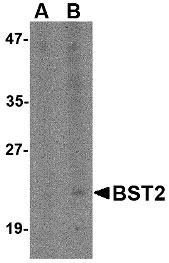
| Application Notes | Bst2 antibody can be used for detection of Bst2 by Western blot at 1 - 2 µg/mL. |
| Gene ID | 69550 |
|---|---|
| Other Names | Bone marrow stromal antigen 2, BST-2, HM1.24 antigen, CD317, Bst2 |
| Target/Specificity | Bst2; |
| Reconstitution & Storage | Bst2 antibody can be stored at 4℃ for three months and -20℃, stable for up to one year. As with all antibodies care should be taken to avoid repeated freeze thaw cycles. Antibodies should not be exposed to prolonged high temperatures. |
| Precautions | Bst2 Antibody is for research use only and not for use in diagnostic or therapeutic procedures. |
For Research Use Only. Not For Use In Diagnostic Procedures.
| Name | Bst2 |
|---|---|
| Function | IFN-induced antiviral host restriction factor which efficiently blocks the release of diverse mammalian enveloped viruses by directly tethering nascent virions to the membranes of infected cells. Acts as a direct physical tether, holding virions to the cell membrane and linking virions to each other. The tethered virions can be internalized by endocytosis and subsequently degraded or they can remain on the cell surface. In either case, their spread as cell-free virions is restricted. Its target viruses belong to diverse families, including retroviridae: human immunodeficiency virus type 1 (HIV-1), mouse mammary tumor virus (MMTV) and murine leukemia virus (MLV), filoviridae: ebola virus (EBOV), arenaviridae: lassa virus (LASV), and rhabdoviridae: vesicular stomatitis virus (VSV). Can inhibit cell surface proteolytic activity of MMP14 causing decreased activation of MMP15 which results in inhibition of cell growth and migration. Can stimulate signaling by LILRA4/ILT7 and consequently provide negative feedback to the production of IFN by plasmacytoid dendritic cells in response to viral infection. Plays a role in the organization of the subapical actin cytoskeleton in polarized epithelial cells. |
| Cellular Location | Golgi apparatus, trans-Golgi network. Cell membrane; Single-pass type II membrane protein. Cell membrane; Lipid-anchor, GPI-anchor. Late endosome. Membrane raft. Cytoplasm Apical cell membrane. Note=Shuttles between the cell membrane, where it is present predominantly in membrane/lipid rafts, and the trans-Golgi network. Forms a complex with MMP14 and localizes to the cytoplasm (By similarity). |
| Tissue Location | In naive mice, specifically expressed on type I interferon-producing cells (at protein level) |
Provided below are standard protocols that you may find useful for product applications.
BACKGROUND
Bst2 Antibody: Bst2 was first identified as a novel terminal B-cell-restricted antigen termed HM1.24 that is expressed on certain bone marrow cells as well as other tissues. The surface expression of Bst2 on fibroblast cell lines facilitated the stromal cell-dependent growth of DW34, a pre-B-cell line, suggesting that this protein is involved in pre-B-cell growth. Later experiments also showed it to be upregulated in several myeloma cells. More recently, Bst2 was identified an inhibitor of retroviral release from human cells whose activity is antagonized by the HIV-1 accessory protein Vpu. While Bst2 caused retention of virions on cell surfaces or endocytosis into Bst2-positive compartments, its depletion abolished the viral requirement for Vpu for virus release. This activity may represent a potential therapeutic strategy for the treatment of HIV/AIDS.
REFERENCES
Goto T, Kennel SJ, Abe M, et al. A novel membrane antigen selectively expressed on terminally differentiated human B cells. Blood1994; 84:1922-30.
Ishikawa J, Kaisho T, Tomizawa H, et al. Molecular cloning and chromosomal mapping of a bone marrow stromal cell surface gene, BST2, that may be involved in pre-B-cell growth. Genomics1995; 26:527-34.
Ohtomo T, Sugamata Y, Ozaki Y, et al. Molecular cloning and characterization of a surface antigen preferentially overexpressed on multiple myeloma cells. Biochem. Biophys. Res. Commun.1999; 258:583-91.
Neil SJ, Zang T, and Bieniasz PD. Tetherin inhibits retrovirus release and is antagonized by HIV-1 Vpu. Nature2008; 451:425-30.

终于等到您。ABCEPTA(百远生物)抗体产品。
点击下方“我要评价 ”按钮提交您的反馈信息,您的反馈和评价是我们最宝贵的财富之一,
我们将在1-3个工作日内处理您的反馈信息。
如有疑问,联系:0512-88856768 tech-china@abcepta.com.


