


 癌症的基本特征包括细胞增殖、血管生成、迁移、凋亡逃避机制和细胞永生等。找到癌症发生过程中这些通路的关键标记物和对应的抗体用于检测至关重要。
 为您推荐一个泛素化位点预测神器——泛素化分析工具,可以为您的蛋白的泛素化位点作出预测和评分。
 细胞自噬受体图形绘图工具为你的蛋白的细胞受体结合位点作出预测和评分,识别结合到自噬通路中的蛋白是非常重要的,便于让我们理解自噬在正常生理、病理过程中的作用,如发育、细胞分化、神经退化性疾病、压力条件下、感染和癌症。
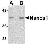
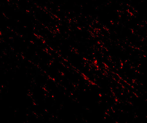
Nanos1 Antibody
- 产品详情
- 实验流程
- 背景知识
Application
| WB, IF, E |
|---|---|
| Primary Accession | Q8WY41 |
| Other Accession | Q8WY41, 41688589 |
| Reactivity | Human, Mouse, Rat |
| Host | Rabbit |
| Clonality | Polyclonal |
| Isotype | IgG |
| Calculated MW | 30230 Da |
| Concentration (mg/ml) | 1 mg/mL |
| Conjugate | Unconjugated |
| Application Notes | Nanos1 antibody can be used for detection of Nanos1 by Western blot at 1 - 2 µg/mL. For immunofluorescence start at 20 µg/mL. |
| Gene ID | 340719 |
|---|---|
| Other Names | Nanos homolog 1, NOS-1, EC_Rep1a, NANOS1, NOS1 |
| Target/Specificity | NANOS1; |
| Reconstitution & Storage | Nanos1 antibody can be stored at 4℃ for three months and -20℃, stable for up to one year. As with all antibodies care should be taken to avoid repeated freeze thaw cycles. Antibodies should not be exposed to prolonged high temperatures. |
| Precautions | Nanos1 Antibody is for research use only and not for use in diagnostic or therapeutic procedures. |
| Name | NANOS1 |
|---|---|
| Synonyms | NOS1 |
| Function | May act as a translational repressor which regulates translation of specific mRNAs by forming a complex with PUM2 that associates with the 3'-UTR of mRNA targets. Capable of interfering with the proadhesive and anti-invasive functions of E-cadherin. Up-regulates the production of MMP14 to promote tumor cell invasion. |
| Cellular Location | Cytoplasm, perinuclear region. Cytoplasm Note=Colocalizes with SNAPIN and PUM2 in the perinuclear region of germ cells. |
| Tissue Location | Testis and ovary (at protein level). Predominantly expressed in testis. Specifically expressed during germline development. In adult tissues, it is mainly expressed in spermatogonia, the stem cells of the germline. Also expressed during meiosis in spermatocytes. Not present in late, post-meiotic stage germ cells Expressed in fetal ovaries, while it is weakly or not expressed in mature postmeiotic oocytes, suggesting that it may be expressed in premeiotic female germ cells. Expressed at high levels only in the E- cadherin deficient cell lines. Highly expressed in lung carcinomas and mostly detected in invasive tumor cells and its expression correlates with tumor aggressiveness. |
For Research Use Only. Not For Use In Diagnostic Procedures.
Provided below are standard protocols that you may find useful for product applications.
BACKGROUND
Nanos1 Antibody: Nanos1 is one of three known mammalian homologs to the Drosophila gene nanos. Nanos1 is an RNA-binding protein containing a zinc-finger motif and is expressed in the developing nervous system and continues in the adult brain. Interestingly, unlike mice deficient in either nanos2 or nanos3, mice lacking the nanos1 gene develop normally with no sign of abnormalities. Recently it has been found that expression of nanos1 mRNA is down-regulated by E-cadherin in a human breast cancer cell line and the amino-terminal domain on Nanos1 interacts with the E-cadherin-binding protein p120ctn. Furthermore, overexpression of Nanos1 in human colorectal DLD1 cancer cells functionally abolished cell-cell adhesion, allowing the cancer cells to develop strong migratory and invasive properties. These results suggest that targeting Nanos1 might prove an effective strategy in the treatment of E-cadherin-negative tumors.
REFERENCES
Jaruleska J, Kotecki M, Kusz K, et al. Conservation of a Pumilio-Nanos complex from Drosophila germ plasm to human germ cells. Dev. Genes Evol.2003; 213:120-6.
Tsuda M, Sasaoka Y, Kiso M, et al. Conserved role of nanos proteins in germ cell development. Science2003; 301:1239-41.
Haraguchi S, Tsuda M, Kitajima S, et al. Nanos1: a mouse nanos gene expressed in the central nervous system is dispensable for normal development. Mech. Dev.2003; 120:721-31.
Strumane K, Bonnomet A, Stove A, et al. E-cadherin regulates human Nanos1, which interacts with p120ctn and induces tumor cell migration and invasion. Cancer Res.2006; 66:10007-15.
终于等到您。ABCEPTA(百远生物)抗体产品。
点击下方“我要评价 ”按钮提交您的反馈信息,您的反馈和评价是我们最宝贵的财富之一,
我们将在1-3个工作日内处理您的反馈信息。
如有疑问,联系:0512-88856768 tech-china@abcepta.com.


