


 癌症的基本特征包括细胞增殖、血管生成、迁移、凋亡逃避机制和细胞永生等。找到癌症发生过程中这些通路的关键标记物和对应的抗体用于检测至关重要。
 为您推荐一个泛素化位点预测神器——泛素化分析工具,可以为您的蛋白的泛素化位点作出预测和评分。
 细胞自噬受体图形绘图工具为你的蛋白的细胞受体结合位点作出预测和评分,识别结合到自噬通路中的蛋白是非常重要的,便于让我们理解自噬在正常生理、病理过程中的作用,如发育、细胞分化、神经退化性疾病、压力条件下、感染和癌症。
GDF6 Antibody
- 产品详情
- 实验流程
- 背景知识
Application
| WB, E |
|---|---|
| Primary Accession | Q6KF10 |
| Other Accession | NP_001001557, 48475062 |
| Reactivity | Human, Mouse, Rat |
| Host | Rabbit |
| Clonality | Polyclonal |
| Isotype | IgG |
| Calculated MW | 50662 Da |
| Concentration (mg/ml) | 1 mg/mL |
| Conjugate | Unconjugated |
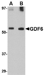
| Application Notes | GDF6 antibody can be used for detection of GDF6 by Western blot at 0.5 to 1 µg/mL. |
| Gene ID | 392255 |
|---|---|
| Other Names | Growth/differentiation factor 6, GDF-6, Bone morphogenetic protein 13, BMP-13, Growth/differentiation factor 16, GDF6, BMP13, GDF16 |
| Target/Specificity | GDF6; |
| Reconstitution & Storage | GDF6 antibody can be stored at 4℃ for three months and -20℃, stable for up to one year. As with all antibodies care should be taken to avoid repeated freeze thaw cycles. Antibodies should not be exposed to prolonged high temperatures. |
| Precautions | GDF6 Antibody is for research use only and not for use in diagnostic or therapeutic procedures. |
| Name | GDF6 |
|---|---|
| Synonyms | BMP13, GDF16 |
| Function | Growth factor that controls proliferation and cellular differentiation in the retina and bone formation. Plays a key role in regulating apoptosis during retinal development. Establishes dorsal- ventral positional information in the retina and controls the formation of the retinotectal map (PubMed:23307924). Required for normal formation of bones and joints in the limbs, skull, digits and axial skeleton. Plays a key role in establishing boundaries between skeletal elements during development. Regulation of GDF6 expression seems to be a mechanism for evolving species-specific changes in skeletal structures. Seems to positively regulate differentiation of chondrogenic tissue through the growth factor receptors subunits BMPR1A, BMPR1B, BMPR2 and ACVR2A, leading to the activation of SMAD1- SMAD5-SMAD8 complex. The regulation of chondrogenic differentiation is inhibited by NOG (PubMed:26643732). Also involved in the induction of adipogenesis from mesenchymal stem cells. This mechanism acts through the growth factor receptors subunits BMPR1A, BMPR2 and ACVR2A and the activation of SMAD1-SMAD5-SMAD8 complex and MAPK14/p38 (By similarity). |
| Cellular Location | Secreted. |
For Research Use Only. Not For Use In Diagnostic Procedures.
Provided below are standard protocols that you may find useful for product applications.
BACKGROUND
GDF6 Antibody: Growth differentiation factors (GDFs) are members of the transforming growth factor (TGF) superfamily that is involved in embryonic development and adult tissue homeostasis. Both GDF6 and GDF7 are closely related to GDF5 which has been shown to induce activation of plasminogen activator, thereby inducing angiogenesis. It is predominantly expressed in long bones during fetal embryonic development and is involved in bone formation. In Xenopus, GDF6 is expressed at the edge of the neural plate and within the anterior neural plate including the eye fields. GDF6 is required for normal formation of some bones and joints in the limbs, skull, and axial skeleton. It may regulate patterning of the ectoderm by interacting with bone morphogenetic proteins (BMPs), and control eye development. Mutations in this gene result in colobomata, which are congenital abnormalities in ocular development, and in Klippel-Feil syndrome (KFS), a congenital disorder of spinal segmentation.
REFERENCES
Massague J. 1990. The transforming growth factor-beta family. Ann. Rev. Cell Biol.6:597-641.
McPherron AC, Lawler AM, and Lee SJ. Regulation of skeletal muscle mass in mice by a new TGF-beta superfamily member. Nature1997; 387:83-90.
Hanel ML and Hensey C. Eye and neural defects associated with loss of GDF6. BMC Dev. Biol.2006; 6:43.
Settle SH Jr., Rountree RB, Sinha A, et al. Multiple joint and skeletal patterning defects caused by single and double mutations in the mouse Gdf6 and Gdf5 genes. Dev. Biol.2003; 254:116-130.
终于等到您。ABCEPTA(百远生物)抗体产品。
点击下方“我要评价 ”按钮提交您的反馈信息,您的反馈和评价是我们最宝贵的财富之一,
我们将在1-3个工作日内处理您的反馈信息。
如有疑问,联系:0512-88856768 tech-china@abcepta.com.


