


 癌症的基本特征包括细胞增殖、血管生成、迁移、凋亡逃避机制和细胞永生等。找到癌症发生过程中这些通路的关键标记物和对应的抗体用于检测至关重要。
 为您推荐一个泛素化位点预测神器——泛素化分析工具,可以为您的蛋白的泛素化位点作出预测和评分。
 细胞自噬受体图形绘图工具为你的蛋白的细胞受体结合位点作出预测和评分,识别结合到自噬通路中的蛋白是非常重要的,便于让我们理解自噬在正常生理、病理过程中的作用,如发育、细胞分化、神经退化性疾病、压力条件下、感染和癌症。
POLR3F Antibody
- 产品详情
- 实验流程
- 背景知识
Application 
| WB, IF, E, IHC-P |
|---|---|
| Primary Accession | Q9H1D9 |
| Other Accession | CAC11110, 10636565 |
| Reactivity | Human, Mouse, Rat |
| Host | Rabbit |
| Clonality | Polyclonal |
| Isotype | IgG |
| Calculated MW | 35684 Da |
| Concentration (mg/ml) | 1 mg/mL |
| Conjugate | Unconjugated |
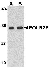
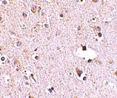
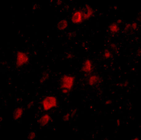
| Application Notes | POLR3F antibody can be used for detection of POLR3F by Western blot at 0.5 - 1 µg/mL. Antibody can also be used for immunohistochemistry starting at 2.5 µg/mL. For immunofluorescence start at 20 µg/mL. |
| Gene ID | 10621 |
|---|---|
| Other Names | DNA-directed RNA polymerase III subunit RPC6, RNA polymerase III subunit C6, DNA-directed RNA polymerase III subunit F, RNA polymerase III 39 kDa subunit, RPC39, POLR3F |
| Target/Specificity | POLR3F; |
| Reconstitution & Storage | POLR3F antibody can be stored at 4℃ for three months and -20℃, stable for up to one year. As with all antibodies care should be taken to avoid repeated freeze thaw cycles. Antibodies should not be exposed to prolonged high temperatures. |
| Precautions | POLR3F Antibody is for research use only and not for use in diagnostic or therapeutic procedures. |
For Research Use Only. Not For Use In Diagnostic Procedures.
| Name | POLR3F (HGNC:15763) |
|---|---|
| Function | DNA-dependent RNA polymerase catalyzes the transcription of DNA into RNA using the four ribonucleoside triphosphates as substrates (PubMed:20413673, PubMed:21358628, PubMed:33558764, PubMed:34675218). Specific peripheric component of RNA polymerase III (Pol III) which synthesizes small non-coding RNAs including 5S rRNA, snRNAs, tRNAs and miRNAs from at least 500 distinct genomic loci. Part of POLR3C/RPC3- POLR3F/RPC6-POLR3G/RPC7 heterotrimer that coordinates the dynamics of Pol III stalk and clamp modules during the transition from apo to elongation state (PubMed:20413673, PubMed:33558764, PubMed:33558766). Pol III plays a key role in sensing and limiting infection by intracellular bacteria and DNA viruses, including varicella zoster virus. Acts as a nuclear and cytosolic DNA sensor detecting AT-rich DNA, involved in innate immune response. Can sense non-self dsDNA that serves as template for transcription into dsRNA. The non-self RNA polymerase III transcripts, such as Epstein-Barr virus-encoded RNAs (EBERs) induce type I interferon and NF-kappa-B through the RIG-I pathway (PubMed:19609254, PubMed:19631370, PubMed:30211253). Preferentially binds double-stranded DNA (dsDNA) (PubMed:21358628). |
| Cellular Location | Nucleus. |
Provided below are standard protocols that you may find useful for product applications.
BACKGROUND
POLR3F Antibody: The human POLR3F is a component of RNA III polymerase. RNA polymerase III transcribes many essential, small, noncoding RNAs, including the 5S rRNAs and tRNAs. While most pol III-transcribed genes are found scattered throughout the linear chromosome maps or in multiple linear clusters, there is increasing evidence that many of these genes prefer to be spatially clustered, often at or near the nucleolus. This association could create an environment that fosters the coregulation of transcription by pol III with transcription of the large ribosomal RNA repeats by RNA polymerase I (pol I) within the nucleolus. Given the high number of pol III-transcribed genes in all eukaryotic genomes, the spatial organization of these genes is likely to affect a large portion of the other genes in a genome. POLR3F has also been recently identified as an HIV dependency factor (HDF), suggesting that POLR3F may be an important drug target in HIV treatment. At least two isoforms of POLR3F are known to exist.
REFERENCES
Hu P, Wu S, Sun Y, et al. Characterization of human RNA polymerase III identifies orthologues for Saccharomyces cerevisiae RNA polymerase III subunits. Mol. Cell Biol.2002; 22:8044-55.
Haeusler RA and Engelke DR. Spatial organization of transcription by RNA polymerase III. Nucleic Acids Res.2006; 34:4826-36. Epub 2006 Sep 13.
Brass AL, Dykxhoorn DM, Benita Y, et al. Identification of host proteins required for HIV infection through a functional genomic screen. Science2008; 319:921-6.

终于等到您。ABCEPTA(百远生物)抗体产品。
点击下方“我要评价 ”按钮提交您的反馈信息,您的反馈和评价是我们最宝贵的财富之一,
我们将在1-3个工作日内处理您的反馈信息。
如有疑问,联系:0512-88856768 tech-china@abcepta.com.


