


 癌症的基本特征包括细胞增殖、血管生成、迁移、凋亡逃避机制和细胞永生等。找到癌症发生过程中这些通路的关键标记物和对应的抗体用于检测至关重要。
 为您推荐一个泛素化位点预测神器——泛素化分析工具,可以为您的蛋白的泛素化位点作出预测和评分。
 细胞自噬受体图形绘图工具为你的蛋白的细胞受体结合位点作出预测和评分,识别结合到自噬通路中的蛋白是非常重要的,便于让我们理解自噬在正常生理、病理过程中的作用,如发育、细胞分化、神经退化性疾病、压力条件下、感染和癌症。
Spred2 Antibody
- 产品详情
- 实验流程
- 背景知识
Application
| WB, E |
|---|---|
| Primary Accession | Q7Z698 |
| Other Accession | NP_861449, 189571669 |
| Reactivity | Human |
| Host | Rabbit |
| Clonality | Polyclonal |
| Isotype | IgG |
| Calculated MW | 47558 Da |
| Concentration (mg/ml) | 1 mg/mL |
| Conjugate | Unconjugated |
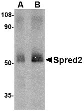
| Application Notes | Spred2 antibody can be used for detection of Spred2 by Western blot at 1 - 2 µg/mL. |
| Gene ID | 200734 |
|---|---|
| Other Names | Sprouty-related, EVH1 domain-containing protein 2, Spred-2, SPRED2 |
| Target/Specificity | SPRED2; This Spred2 antibody is predicted to have no cross-reactivity to Spred1 or Spred3. |
| Reconstitution & Storage | Spred2 antibody can be stored at 4℃ for three months and -20℃, stable for up to one year. As with all antibodies care should be taken to avoid repeated freeze thaw cycles. Antibodies should not be exposed to prolonged high temperatures. |
| Precautions | Spred2 Antibody is for research use only and not for use in diagnostic or therapeutic procedures. |
| Name | SPRED2 |
|---|---|
| Function | Negatively regulates Ras signaling pathways and downstream activation of MAP kinases (PubMed:15683364, PubMed:34626534). Recruits and translocates NF1 to the cell membrane, thereby enabling NF1- dependent hydrolysis of active GTP-bound Ras to inactive GDP-bound Ras (PubMed:34626534). Inhibits fibroblast growth factor (FGF)-induced retinal lens fiber differentiation, probably by inhibiting FGF-mediated phosphorylation of ERK1/2 (By similarity). Inhibits TGFB-induced epithelial-to-mesenchymal transition in lens epithelial cells (By similarity). |
| Cellular Location | Cell membrane; Peripheral membrane protein {ECO:0000250|UniProtKB:Q924S7}; Cytoplasmic side {ECO:0000250|UniProtKB:Q924S7}. Cytoplasmic vesicle, secretory vesicle membrane; Peripheral membrane protein; Cytoplasmic side. Cytoplasm. Note=Detected in the cytoplasm of the stratum spinosum cells, where it is associated with cytoplasmic vesicles that are supposed to be secretory granules |
| Tissue Location | Expressed in liver, skin, small intestine, salivary gland and prostate. |
For Research Use Only. Not For Use In Diagnostic Procedures.
Provided below are standard protocols that you may find useful for product applications.
BACKGROUND
Spred2 Antibody: Spred2 is a member of the Sprouty family, a group of proteins that act as negative regulators during development. Like Spred1, Spred2 acts by suppressing the phosphorylation and activation of Raf. The Spred proteins have also been implicated in the negative feedback regulation of FGF signaling in embryogenesis and angiogenesis. Further studies have shown that expression levels of Spred1 and Spred2 proteins are inversely correlated with the incidence of tumor invasion and metastasis in human hepatocellular carcinoma (HHC), suggesting that these proteins could be useful as prognostic factors and therapeutic targets in HCC. At least two isoforms of Spred2 are known to exist.
REFERENCES
Wakioka T, Sasaki A, Kato R, et al. Spred is a Sprouty-related suppressor of Ras signalling. Nature2001; 412:647-51.
Casci T, Vinos J, and Freeman M. Sprouty, an intracellular inhibitor of Ras signaling. Cell1999; 96:655-65.
Minowada G, Jarvis LA, Chi CL, et al. Vertebrate Sprouty genes are induced by FGF signaling and can cause chondrodysplasia when overexpressed. Development1999; 126:4465-75.
Lee SH, Schloss DJ, Jarvis L, et al. Inhibition of angiogenesis by a mouse sprouty protein. J. Biol. Chem.2001; 276:4128-33.
终于等到您。ABCEPTA(百远生物)抗体产品。
点击下方“我要评价 ”按钮提交您的反馈信息,您的反馈和评价是我们最宝贵的财富之一,
我们将在1-3个工作日内处理您的反馈信息。
如有疑问,联系:0512-88856768 tech-china@abcepta.com.


