


 癌症的基本特征包括细胞增殖、血管生成、迁移、凋亡逃避机制和细胞永生等。找到癌症发生过程中这些通路的关键标记物和对应的抗体用于检测至关重要。
 为您推荐一个泛素化位点预测神器——泛素化分析工具,可以为您的蛋白的泛素化位点作出预测和评分。
 细胞自噬受体图形绘图工具为你的蛋白的细胞受体结合位点作出预测和评分,识别结合到自噬通路中的蛋白是非常重要的,便于让我们理解自噬在正常生理、病理过程中的作用,如发育、细胞分化、神经退化性疾病、压力条件下、感染和癌症。
TTBK2 Antibody
- 产品详情
- 实验流程
- 背景知识
Application 
| E, IHC-P |
|---|---|
| Primary Accession | Q6IQ55 |
| Other Accession | AAH71556, 47940064 |
| Reactivity | Human |
| Host | Rabbit |
| Clonality | Polyclonal |
| Isotype | IgG |
| Calculated MW | 137412 Da |
| Concentration (mg/ml) | 1 mg/mL |
| Conjugate | Unconjugated |
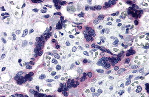
| Application Notes | TTBK2 antibody can be used for detection of TTBK2 by immunohistochemistry at 5 µg/mL. |
| Gene ID | 146057 |
|---|---|
| Other Names | Tau-tubulin kinase 2, 2.7.11.1, TTBK2, KIAA0847 |
| Target/Specificity | TTBK2; TTBK2 antibody is human specific. At least four isoforms of TTBK2 are known to exist; this antibody will detect only the two largest isoforms. |
| Reconstitution & Storage | TTBK2 antibody can be stored at 4℃ for three months and -20℃, stable for up to one year. As with all antibodies care should be taken to avoid repeated freeze thaw cycles. Antibodies should not be exposed to prolonged high temperatures. |
| Precautions | TTBK2 Antibody is for research use only and not for use in diagnostic or therapeutic procedures. |
For Research Use Only. Not For Use In Diagnostic Procedures.
| Name | TTBK2 |
|---|---|
| Synonyms | KIAA0847 |
| Function | Serine/threonine kinase that acts as a key regulator of ciliogenesis: controls the initiation of ciliogenesis by binding to the distal end of the basal body and promoting the removal of CCP110, which caps the mother centriole, leading to the recruitment of IFT proteins, which build the ciliary axoneme. Has some substrate preference for proteins that are already phosphorylated on a Tyr residue at the +2 position relative to the phosphorylation site. Able to phosphorylate tau on serines in vitro (PubMed:23141541). Phosphorylates MPHOSPH9 which promotes its ubiquitination and proteasomal degradation, loss of MPHOSPH9 facilitates the removal of the CP110-CEP97 complex (a negative regulator of ciliogenesis) from the mother centrioles, promoting the initiation of ciliogenesis (PubMed:30375385). Required for recruitment of CPLANE2 and INTU to the mother centriole (By similarity). |
| Cellular Location | Cell projection, cilium. Cytoplasm, cytoskeleton, cilium basal body. Cytoplasm, cytoskeleton, microtubule organizing center, centrosome, centriole. Cytoplasm, cytosol. Nucleus Note=Localizes at the transition zone, a region between the basal body and the ciliary axoneme (PubMed:23141541). May also be present in cytosol and, at lower level in the nucleus (PubMed:21548880) |
Provided below are standard protocols that you may find useful for product applications.
BACKGROUND
TTBK2 Antibody: Tau tubulin kinase (TTBK2) is a serine/threonine kinase whose substrates are thought to include the tau and tubulin proteins. Mutations in the TTBK2 gene cause spinocerebellar ataxia type 11 (SCA11), a neurodegenerative disease characterized by progressive ataxia and atrophy of the cerebellum and brainstem. Recent studies have shown that increased levels of TTBK2 expression in kidney carcinoma and melanoma cell lines increases resistance to the chemotherapeutic agent Sunitinib, and that reduction of TTBK2 though the use of siRNA sensitized these cell lines to the effects of Sunitib, suggesting that TTBK2 can be used as a resistance marker as well a potential target to overcome resistance.
REFERENCES
Kitano-Takahashi M, Morita H, Kondo S, et al. Expression, purification and crystallization of a human tau-tubulin kinase 2 that phosphorylates tau protein. Acta Crystallogr. Sect. F Struct. Biol. Cryst. Commun. 2007; 63:602-4.
Edener U, Kurth I, Meiner A, et al. Missense exchanges in the TTBK2 gene mutated in SCA11. J. Neurol. 2009; 1856-9.
Bender C and Ullrich A. PRKX, TTBK2 and RSK4 expression causes sunitinib resistance in kidney carcinoma- and melanoma cell lines. Int. J. Cancer 2011; epub.

终于等到您。ABCEPTA(百远生物)抗体产品。
点击下方“我要评价 ”按钮提交您的反馈信息,您的反馈和评价是我们最宝贵的财富之一,
我们将在1-3个工作日内处理您的反馈信息。
如有疑问,联系:0512-88856768 tech-china@abcepta.com.


